Explore
 Validate
 Learn
Learn Western blot
 Immunocytochemistry
Antibody data
- Antibody Data
- Antigen structure
- References [3]
- Comments [0]
- Validations
- Immunocytochemistry [1]
- Immunohistochemistry [1]
Submit
Validation data
Reference
Comment
Report error
- Product number
- HPA011155 - Provider product page

- Provider
- Atlas Antibodies
- Proper citation
- Atlas Antibodies Cat#HPA011155, RRID:AB_1852685
- Product name
- Anti-LAIR1
- Antibody type
- Polyclonal
- Description
- Polyclonal Antibody against Human LAIR1, Gene description: leukocyte-associated immunoglobulin-like receptor 1, Alternative Gene Names: CD305, Validated applications: ICC, IHC, WB, Uniprot ID: Q6GTX8, Storage: Store at +4°C for short term storage. Long time storage is recommended at -20°C.
- Reactivity
- Human
- Host
- Rabbit
- Conjugate
- Unconjugated
- Isotype
- IgG
- Vial size
- 100 µl
- Concentration
- 0.4 mg/ml
- Storage
- Store at +4°C for short term storage. Long time storage is recommended at -20°C.
- Handling
- The antibody solution should be gently mixed before use.
Submitted references Single-cell RNA sequencing shows the immunosuppressive landscape and tumor heterogeneity of HBV-associated hepatocellular carcinoma
Cancer immunotherapy by NC410, a LAIR-2 Fc protein blocking human LAIR-collagen interaction.
Evidence for C1q-mediated crosslinking of CD33/LAIR-1 inhibitory immunoreceptors and biological control of CD33/LAIR-1 expression
Ho D, Tsui Y, Chan L, Sze K, Zhang X, Cheu J, Chiu Y, Lee J, Chan A, Cheung E, Yau D, Chia N, Lo I, Sham P, Cheung T, Wong C, Ng I
Nature Communications 2021;12(1)
Nature Communications 2021;12(1)
Cancer immunotherapy by NC410, a LAIR-2 Fc protein blocking human LAIR-collagen interaction.
Ramos MIP, Tian L, de Ruiter EJ, Song C, Paucarmayta A, Singh A, Elshof E, Vijver SV, Shaik J, Bosiacki J, Cusumano Z, Jensen C, Willumsen N, Karsdal MA, Liu L, Langermann S, Willems S, Flies D, Meyaard L
eLife 2021 Jun 14;10
eLife 2021 Jun 14;10
Evidence for C1q-mediated crosslinking of CD33/LAIR-1 inhibitory immunoreceptors and biological control of CD33/LAIR-1 expression
Son M, Diamond B, Volpe B, Aranow C, Mackay M, Santiago-Schwarz F
Scientific Reports 2017;7(1)
Scientific Reports 2017;7(1)
No comments: Submit comment
Supportive validation
- Submitted by
- Atlas Antibodies (provider)
- Main image
- Experimental details
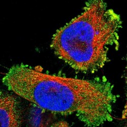
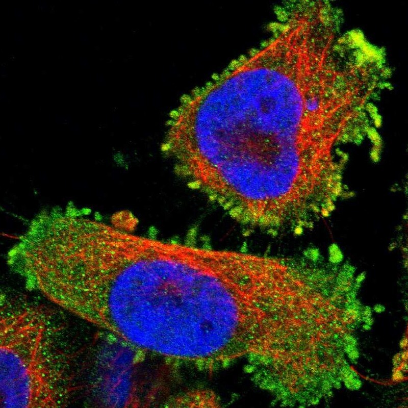
- Immunofluorescent staining of human cell line U-251 MG shows localization to plasma membrane.
- Sample type
- Human
Supportive validation
- Submitted by
- Atlas Antibodies (provider)
- Enhanced method
- Orthogonal validation
- Main image
- Experimental details
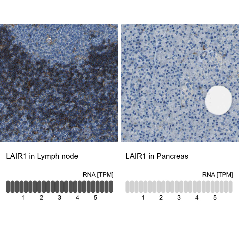
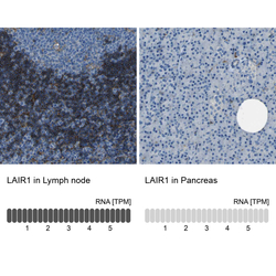
- Immunohistochemistry analysis in human lymph node and pancreas tissues using HPA011155 antibody. Corresponding LAIR1 RNA-seq data are presented for the same tissues.
- Sample type
- Human
- Protocol
- Protocol