Explore
 Validate
 Learn
LearnACC-001-200UL
antibody from Invitrogen Antibodies
Targeting: CACNA1A
APCA, CACNL1A4, Cav2.1, EA2, FHM, HPCA, MHP, MHP1, SCA6
 Western blot
Antibody data
- Antibody Data
- Antigen structure
- References [0]
- Comments [0]
- Validations
- Western blot [1]
- Immunohistochemistry [1]
Submit
Validation data
Reference
Comment
Report error
- Product number
- ACC-001-200UL - Provider product page

- Provider
- Invitrogen Antibodies
- Product name
- CACNA1A (CaV2.1) Polyclonal Antibody
- Antibody type
- Polyclonal
- Antigen
- Other
- Reactivity
- Human, Mouse, Rat
- Host
- Rabbit
- Isotype
- IgG
- Vial size
- 200 µL
- Concentration
- 0.8 mg/mL
- Storage
- -20° C, Avoid Freeze/Thaw Cycles
No comments: Submit comment
Supportive validation
- Submitted by
- Invitrogen Antibodies (provider)
- Main image
- Experimental details
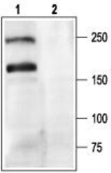
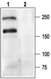
- Western blot analysis of rat brain membranes: - 1. Anti-CACNA1A (CaV2.1) Antibody (#ACC-001), (1:200). 2. Anti-CACNA1A (CaV2.1) Antibody , preincubated with CACNA1A/Cav2.1 Blocking Peptide (#BLP-CC001).
Supportive validation
- Submitted by
- Invitrogen Antibodies (provider)
- Main image
- Experimental details
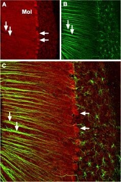
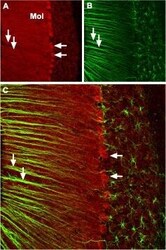
- Expression of CACNA1A in mouse cerebellum - Immunohistochemical staining ofmouse cerebellum with Anti-CACNA1A (CaV2.1) Antibody (#ACC-001), (1:100). A. CACNA1Achannel (red) appears in Purkinje cells (horizontal arrows) and is distributed diffusely in the molecular layer (Mol) including in astrocytic fibers (vertical arrows). B. Staining of astrocytic fibers with glial fibrillary acidic protein in the section demonstrates the location of astrocytic fibers in the molecular layer. C. Merged image of panels A and B.