Explore
 Validate
 Learn
Learn Western blot
 Immunocytochemistry
 Immunoprecipitation
Antibody data
- Antibody Data
- Antigen structure
- References [1]
- Comments [0]
- Validations
- Immunocytochemistry [4]
- Other assay [1]
Submit
Validation data
Reference
Comment
Report error
- Product number
- PA5-17382 - Provider product page

- Provider
- Invitrogen Antibodies
- Product name
- HSP40 Polyclonal Antibody
- Antibody type
- Polyclonal
- Antigen
- Synthetic peptide
- Description
- It is not recommended to aliquot this antibody. This antibody is not cross-reactive with other HSPs.
- Reactivity
- Human, Mouse, Rat
- Host
- Rabbit
- Isotype
- IgG
- Vial size
- 100 μL
- Concentration
- 35 μg/mL
- Storage
- -20°C
Submitted references The interplay between redox signalling and proteostasis in neurodegeneration: In vivo effects of a mitochondria-targeted antioxidant in Huntington's disease mice.
Pinho BR, Duarte AI, Canas PM, Moreira PI, Murphy MP, Oliveira JMA
Free radical biology & medicine 2020 Jan;146:372-382
Free radical biology & medicine 2020 Jan;146:372-382
No comments: Submit comment
Supportive validation
- Submitted by
- Invitrogen Antibodies (provider)
- Main image
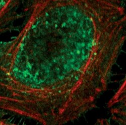
- Experimental details
- Immunofluorescent analysis of HSP40 in HeLa cells, heat-treated, using a HSP40 polyclonal antibody (Product # PA5-17382) (green). Actin filaments are labeled with a fluorescent red phalloidin.

- Submitted by
- Invitrogen Antibodies (provider)
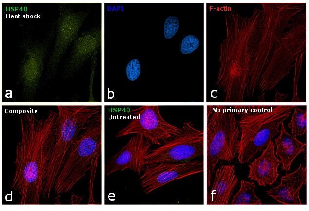
- Main image
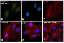
- Experimental details
- Immunofluorescence analysis of HSP40 was performed using 70% confluent log phase HeLa cells heat shocked at 43°C for 1 hr. The cells were fixed with 4% paraformaldehyde for 10 minutes, permeabilized with 0.1% Triton™ X-100 for 15 minutes, and blocked with 1% BSA for 1 hour at room temperature. The cells were labeled with HSP40 Polyclonal Antibody (Product # PA5-17382) at 1:400 dilution in 0.1% BSA, incubated at 4 degree Celsius overnight and then labeled with Goat anti-Rabbit IgG (H+L) Superclonal™ Secondary Antibody, Alexa Fluor® 488 conjugate (Product # A27034) at a dilution of 1:2000 for 45 minutes at room temperature (Panel a: green). Nuclei (Panel b: blue) were stained with ProLong™ Diamond Antifade Mountant with DAPI (Product # P36962). F-actin (Panel c: red) was stained with Rhodamine Phalloidin (Product # R415, 1:300). Panel d represents the merged image showing nuclear translocation upon heat shock. Panel e shows untreated HeLa cells with cytoplasmic localization. Panel f represents control cells with no primary to assess background. The images were captured at 60X magnification.

- Submitted by
- Invitrogen Antibodies (provider)
- Main image

- Experimental details
- Immunofluorescent analysis of HSP40 in HeLa cells, heat-treated, using a HSP40 polyclonal antibody (Product # PA5-17382) (green). Actin filaments are labeled with a fluorescent red phalloidin.

- Submitted by
- Invitrogen Antibodies (provider)
- Main image

- Experimental details
- Immunofluorescence analysis of HSP40 was performed using 70% confluent log phase HeLa cells heat shocked at 43°C for 1 hr. The cells were fixed with 4% paraformaldehyde for 10 minutes, permeabilized with 0.1% Triton™ X-100 for 15 minutes, and blocked with 1% BSA for 1 hour at room temperature. The cells were labeled with HSP40 Polyclonal Antibody (Product # PA5-17382) at 1:400 dilution in 0.1% BSA, incubated at 4 degree Celsius overnight and then labeled with Goat anti-Rabbit IgG (Heavy Chain) Superclonal™ Secondary Antibody, Alexa Fluor® 488 conjugate (Product # A27034) at a dilution of 1:2000 for 45 minutes at room temperature (Panel a: green). Nuclei (Panel b: blue) were stained with ProLong™ Diamond Antifade Mountant with DAPI (Product # P36962). F-actin (Panel c: red) was stained with Rhodamine Phalloidin (Product # R415, 1:300). Panel d represents the merged image showing nuclear translocation upon heat shock. Panel e shows untreated HeLa cells with cytoplasmic localization. Panel f represents control cells with no primary to assess background. The images were captured at 60X magnification.
Supportive validation
- Submitted by
- Invitrogen Antibodies (provider)
- Main image

- Experimental details
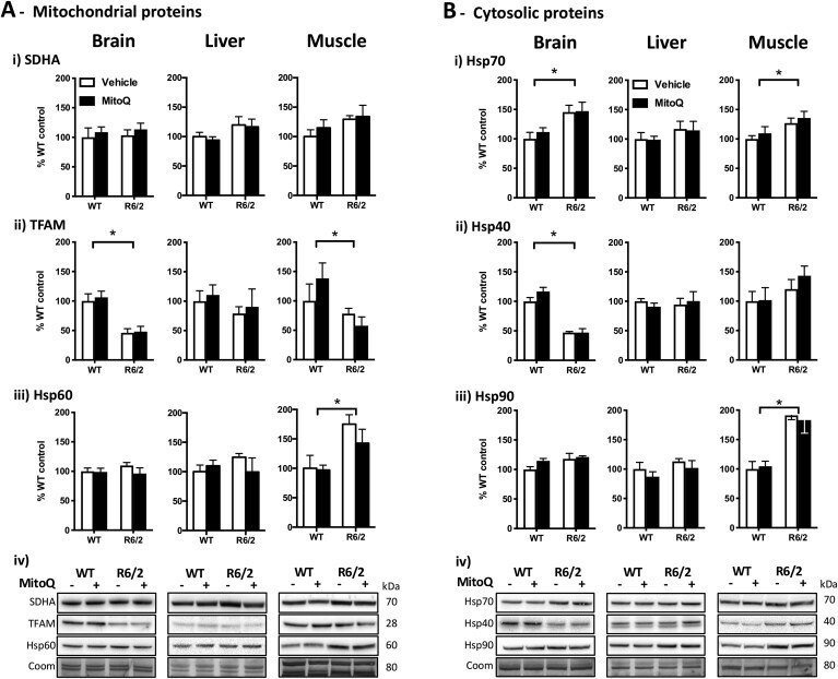
- Fig. 3 Levels of mitochondrial biomarkers and chaperones in WT and R6/2 mice. Western blot quantifications in brain, liver and muscle samples from WT and R6/2 mice treated with vehicle or MitoQ. (A) Mitochondrial biomarkers: i ) SDHA, succinate dehydrogenase complex, subunit A; ii ) TFAM, mitochondrial transcription factor A, iii ) Hsp60; and iv ) representative blots. (B) Cytosolic chaperones: i ) Hsp70; ii ) Hsp40; iii ) Hsp90; and iv ) representative blots. Coom - Coomassie staining loading control. Data are in percentage of the mean of WT-vehicle (control); n = 5; * P < 0.05, genotype effect. Fig. 3