Explore
 Validate
 Learn
LearnPA5-23381
antibody from Invitrogen Antibodies
Targeting: STING1
ERIS, FLJ38577, MITA, MPYS, NET23, STING, TMEM173
 Western blot
 Immunocytochemistry
Antibody data
- Antibody Data
- Antigen structure
- References [3]
- Comments [0]
- Validations
- Immunocytochemistry [4]
- Immunohistochemistry [2]
- Flow cytometry [3]
- Other assay [3]
Submit
Validation data
Reference
Comment
Report error
- Product number
- PA5-23381 - Provider product page

- Provider
- Invitrogen Antibodies
- Product name
- STING Polyclonal Antibody
- Antibody type
- Polyclonal
- Antigen
- Synthetic peptide
- Description
- Sequence homology: Opossum/Zebrafish (83%), Xenopus (72%), Rat (88%).
- Reactivity
- Human, Mouse
- Host
- Rabbit
- Isotype
- IgG
- Vial size
- 100 μg
- Concentration
- 1.0 mg/mL
- Storage
- Store at 4°C short term. For long term storage, store at -20°C, avoiding freeze/thaw cycles.
Submitted references Human rhinovirus promotes STING trafficking to replication organelles to promote viral replication.
ASA404, a vascular disrupting agent, as an experimental treatment approach for brain tumors.
AIM2-Like Receptors Positively and Negatively Regulate the Interferon Response Induced by Cytosolic DNA.
Triantafilou M, Ramanjulu J, Booty LM, Jimenez-Duran G, Keles H, Saunders K, Nevins N, Koppe E, Modis LK, Pesiridis GS, Bertin J, Triantafilou K
Nature communications 2022 Mar 17;13(1):1406
Nature communications 2022 Mar 17;13(1):1406
ASA404, a vascular disrupting agent, as an experimental treatment approach for brain tumors.
Bähr O, Gross S, Harter PN, Kirches E, Mawrin C, Steinbach JP, Mittelbronn M
Oncology letters 2017 Nov;14(5):5443-5451
Oncology letters 2017 Nov;14(5):5443-5451
AIM2-Like Receptors Positively and Negatively Regulate the Interferon Response Induced by Cytosolic DNA.
Nakaya Y, Lilue J, Stavrou S, Moran EA, Ross SR
mBio 2017 Jul 5;8(4)
mBio 2017 Jul 5;8(4)
No comments: Submit comment
Supportive validation
- Submitted by
- Invitrogen Antibodies (provider)
- Main image

- Experimental details
- Immunocytochemistry analysis of STING in RH-30 cells fixed for 10 minutes using 10% formalin and then permeabilized for 5 minutes using 1X PBS + 0.05% Triton X-100. Samples were incubated in STING polyclonal antibody (Product # PA5-23381) using a dilution of 2 µg/mL overnight at 4 °C followed by anti-rabbit DyLight 488 (Green) with a dilution of 1:500. Alpha Tubulin Antibody (DM1A) was used as a co-stain at a 1:1000 dilution and detected with an anti-mouse DyLight 550 (Red) at a 1:500 dilution. Nuclei were counterstained with DAPI (Blue). Cells were imaged using a 40X objective.
- Submitted by
- Invitrogen Antibodies (provider)
- Main image

- Experimental details
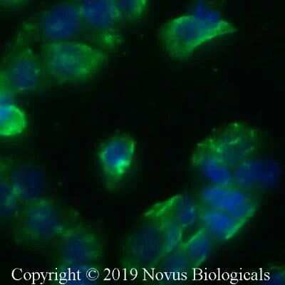
- Immunocytochemistry analysis of STING in HT-29 cells fixed for 10 minutes using 10% formalin and then permeabilized for 5 minutes using 1X PBS + 0.05% Triton-X100. Samples were incubated in STING polyclonal antibody (Product # PA5-23381) using a dilution of 10 µg/mL overnight at 4 °C followed by anti-rabbit DyLight 488 (Green) with a dilution of 1:500. Nuclei were counterstained with DAPI (Blue). Cells were imaged using a 40X objective.
- Submitted by
- Invitrogen Antibodies (provider)
- Main image
- Experimental details
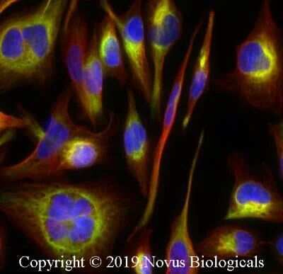
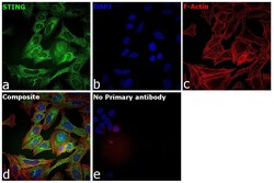
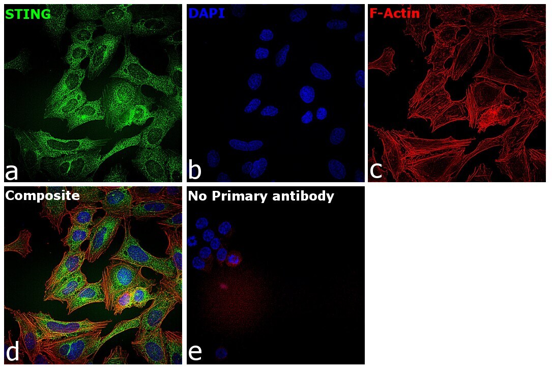
- Immunofluorescence analysis of TMEM173 was performed using 70% confluent log phase U-2 OS cells. The cells were fixed with 4% paraformaldehyde for 10 minutes, permeabilized with 0.01% Triton™ X-100 for 15 minutes, and blocked with 2% BSA for 45 minutes at room temperature. The cells were labeled with TMEM173 (Product # PA5-23381 ) at 1:100 in 0.1% BSA, incubated at 4 degree celsius overnight and then labeled with Donkey anti-Rabbit IgG (H+L) Highly Cross-Adsorbed Secondary Antibody, Alexa Fluor Plus 488 (Product # A32790), (1:2000), for 45 minutes at room temperature (Panel a: Green). Nuclei (Panel b:Blue) were stained with Hoechst 33342 (Product # H1399). F-actin (Panel c: Red) was stained with Rhodamine Phalloidin (Product # R415, 1:300). Panel d represents the merged image predominantly showing endoplasmic reticulum localization. Panel e represents control cells with no primary antibody to assess background. The images were captured at 40X magnification in CellInsight CX7 LZR High-Content Screening (HCS) Platform (Product # CX7C1115LZR). magnification.
- Submitted by
- Invitrogen Antibodies (provider)
- Main image

- Experimental details
- Immunofluorescence analysis of TMEM173 was performed using 70% confluent log phase U-2 OS cells. The cells were fixed with 4% paraformaldehyde for 10 minutes, permeabilized with 0.01% Triton™ X-100 for 15 minutes, and blocked with 2% BSA for 45 minutes at room temperature. The cells were labeled with TMEM173 (Product # PA5-23381 ) at 1:100 in 0.1% BSA, incubated at 4 degree celsius overnight and then labeled with Donkey anti-Rabbit IgG (H+L) Highly Cross-Adsorbed Secondary Antibody, Alexa Fluor Plus 488 (Product # A32790), (1:2000), for 45 minutes at room temperature (Panel a: Green). Nuclei (Panel b:Blue) were stained with Hoechst 33342 (Product # H1399). F-actin (Panel c: Red) was stained with Rhodamine Phalloidin (Product # R415, 1:300). Panel d represents the merged image predominantly showing endoplasmic reticulum localization. Panel e represents control cells with no primary antibody to assess background. The images were captured at 40X magnification in CellInsight CX7 LZR High-Content Screening (HCS) Platform (Product # CX7C1115LZR). magnification.

Supportive validation
- Submitted by
- Invitrogen Antibodies (provider)
- Main image

- Experimental details
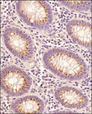
- Immunohistochemical analysis of STING in Human colon cancer tissue section. Samples were incubated in STING polyclonal antibody (Product # PA5-23381) using a dilution of 1:100 followed by HRP-conjugated secondary antibody. The signal was developed using DAB reagent and the nuclei were counterstained with hematoxylin. The antibody generated very weak cytoplasmic staining in columnar epithelial cells with a very strong signal in the secretory/goblet cells.
- Submitted by
- Invitrogen Antibodies (provider)
- Main image
- Experimental details
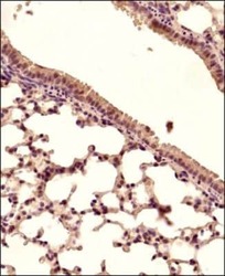
- Immunohistochemical analysis of STING in Mouse lung tissue section. Samples were incubated in STING polyclonal antibody (Product # PA5-23381) using a dilution of 1:150 followed by a HRP-conjugated secondary antibody. The signal was developed using DAB reagent and the nuclei were counterstained with hematoxylin. The antibody generated mainly a cytoplasmic staining in the bronchiolar and alveolar epithelial cells.

Supportive validation
- Submitted by
- Invitrogen Antibodies (provider)
- Main image
- Experimental details
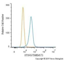
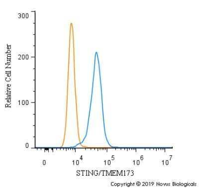
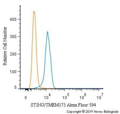
- Flow cytometry of STING in RH30 cells (blue) and a matched isotype control (orange). Samples were incubated in STING polyclonal antibody (Product # PA5-23381) using a dilution of 2.5 µg/mL for 30 minutes at room temperature followed by a Rabbit IgG (H+L) Cross-Adsorbed secondary antibody. Cells were fixed with 4% PFA and then permeabilized with 0.1% saponin.
- Submitted by
- Invitrogen Antibodies (provider)
- Main image
- Experimental details
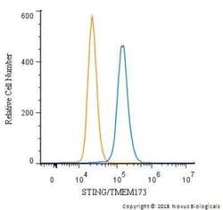
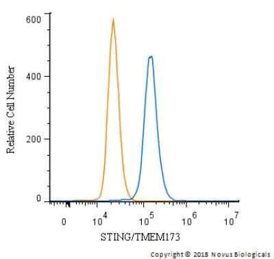
- Flow cytometry of STING in THP-1 cells and a matched isotype control. Samples were incubated in STING polyclonal antibody (Product # PA5-23381) using a dilution of 2.5 µg/mL for 30 minutes at room temperature followed by a Rabbit IgG (H+L) Cross-Adsorbed secondary antibody. Cells were fixed with 4% PFA and then permeabilized with 0.1% saponin.
- Submitted by
- Invitrogen Antibodies (provider)
- Main image
- Experimental details
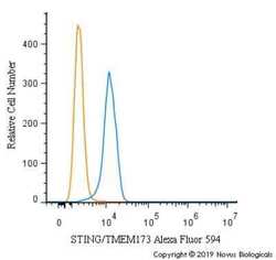
- Flow cytometry of STING in U937 cells. Samples were incubated in STING polyclonal antibody (Product # PA5-23381) using a dilution of 2.5 µg/mL for 30 minutes at room temperature. Antibody (blue) and a matched isotype control (orange). Cells were fixed with 4% PFA and then permeabilized with 0.1% saponin. Both antibodies were conjugated to Alexa Fluor 594.
Supportive validation
- Submitted by
- Invitrogen Antibodies (provider)
- Main image
- Experimental details
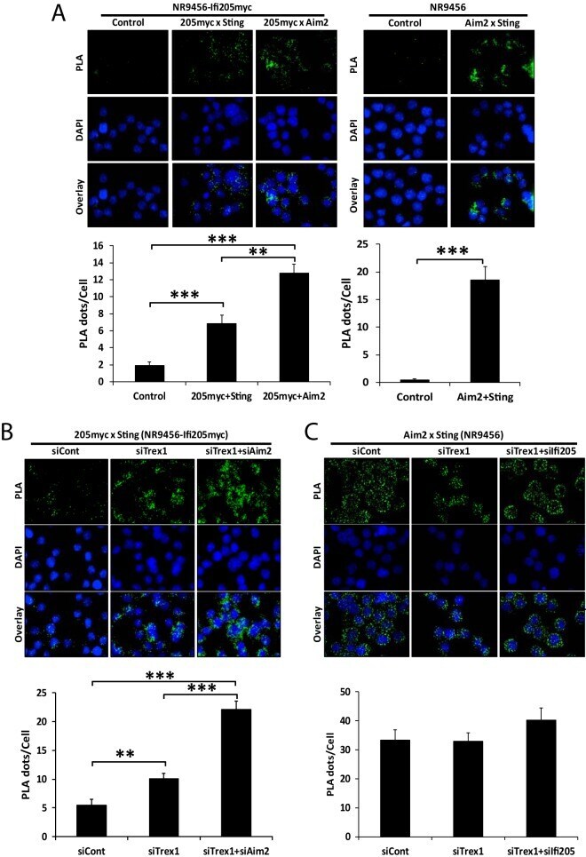
- FIG 7 IFI205-STING interaction increases upon TREX1 and AIM2 depletion. (A) PLA for interactions of IFI205myc-AIM2, IFI205myc-STING, and AIM2-STING in NR9456-IFI205myc or NR9456 cells. The number of cells and images quantified for each condition in NR9456-IFI205myc cells are as follows: IFI205myc-Sting, 141 cells and 10 images; IFI205myc-Aim2, 101 cells and 6 images; control, 65 cells and 5 images. The number of cells and images quantified for each condition in NR9456 cells are as follows: control, 74 cells and 5 images; Aim2-Sting, 47 cells and 5 images. Controls for knockdowns are shown in Fig. S9A . (B) PLA for IFI205myc-STING interactions in NR9456-IFI205myc cells with knockdown of the genes indicated. Quantification: siCont, 102 cells and 5 images; siTrex1, 190 cells and 7 images; siTrex1+siAim2, 108 cells and 5 images. A representative image is shown. This experiment was repeated twice and gave similar results both times. (C) PLA for AIM2-STING interaction in NR9456 cells after knockdown of the genes indicated. Quantification: siCont, 84 cells and 4 images; siTrex1, 33 cells and 4 images; siTrex1+si205, 58 cells and 4 images. PLA dots were quantified and normalized to cell numbers based on DAPI staining with ImageJ software. The values shown are the mean +- the standard error of the mean of different pictures. **, P < 0.005; ***, P < 0.0005. (two-tailed t test). siCont, control siRNA.
- Submitted by
- Invitrogen Antibodies (provider)
- Main image
- Experimental details
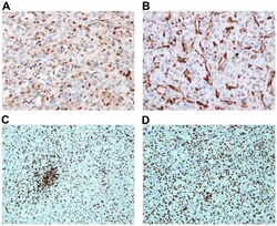
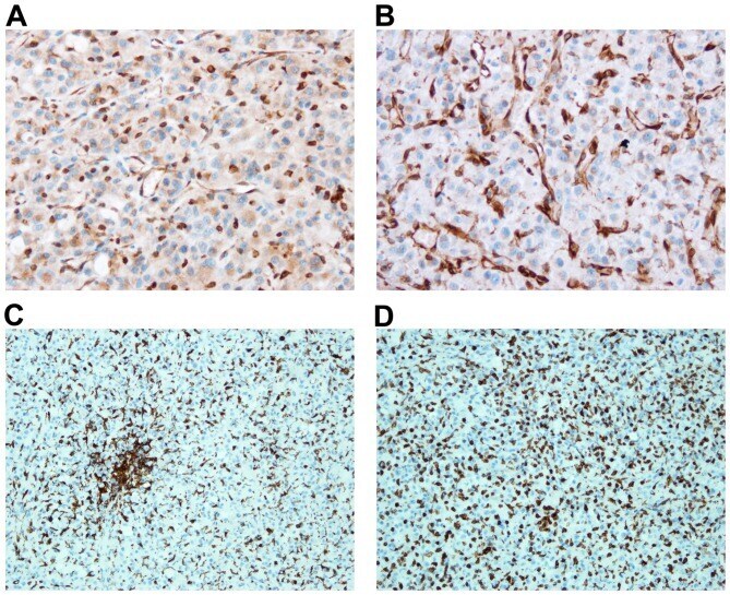
- Figure 8. STING expression and iba1 positive cells in subcutaneous and intracranial U-87 tumors. Immunohistochemistry for STING is shown for subcutaneous (A) and intracranial (B) U-87 tumors. Cells positive for iba1 are stained in subcutaneous (C) and intracranial (D) U-87 tumors.
- Submitted by
- Invitrogen Antibodies (provider)
- Main image
- Experimental details
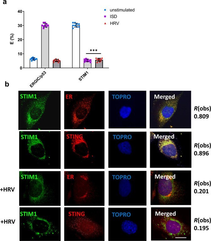
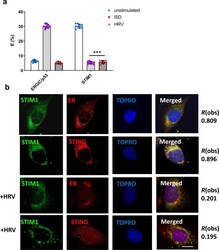
- Fig. 5 STIM1 releases STING upon HRV infection. FRET studies measuring donor (STING) and acceptor (ERGIC or STIM1) interactions in BEAS-2B cells infected with HRV for 2 h or exposed to ISD (1 ug) ( a ). Confocal microscopy of BEAS-2B cells infected with HRV for 2 h assessing colocalization between STIM1, the ER and STING. Data in a is means +/- SD ( n = 3, independent experiments). Statistical significance between unstimulated and virus-infected conditions was assessed by unpaired student's t -test. *** p < 0.001 Data in b is representative of three biological donors with at least 20 technical replicates. Degree of colocalization is presented at R(obs). Bars shown at 10 um.