Explore
 Validate
 Learn
Learn Western blot
Antibody data
- Antibody Data
- Antigen structure
- References [0]
- Comments [0]
- Validations
- Western blot [1]
- Immunocytochemistry [1]
- Immunohistochemistry [2]
- Flow cytometry [1]
Submit
Validation data
Reference
Comment
Report error
- Product number
- AMT-004-25UL - Provider product page

- Provider
- Invitrogen Antibodies
- Product name
- Serotonin Transporter (SERT) (extracellular) Polyclonal Antibody
- Antibody type
- Polyclonal
- Antigen
- Other
- Reactivity
- Human, Mouse, Rat
- Host
- Rabbit
- Isotype
- IgG
- Vial size
- 25 µL
- Concentration
- 0.8 mg/mL
- Storage
- -20° C, Avoid Freeze/Thaw Cycles
No comments: Submit comment
Supportive validation
- Submitted by
- Invitrogen Antibodies (provider)
- Main image

- Experimental details
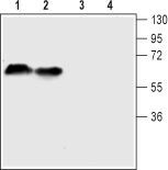
- Western blot analysis of mouse (lanes 1 and 3) and rat (lanes 2 and 4) brain lysates: - 1-2. Anti-Serotonin Transporter (SERT) (extracellular) Antibody (#AMT-004), (1:200).3-4. Anti-Serotonin Transporter (SERT) (extracellular) Antibody , preincubated with Serotonin Transporter/SERT (extracellular) Blocking Peptide (#BLP-MT004).
Supportive validation
- Submitted by
- Invitrogen Antibodies (provider)
- Main image

- Experimental details
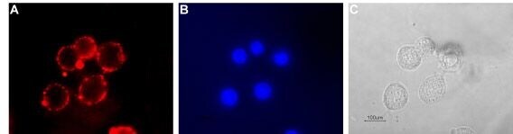
- Expression of serotonin transporter in rat PC12 cells - Cell surface detection of serotonin transporter in live intact rat pheochromocytoma PC12 cells. A. Cells were stained with Anti-Serotonin Transporter (SERT) (extracellular) Antibody (#AMT-004), (1:100), followed by goat Anti-rabbit-AlexaFluor-594 secondary Antibody (red). B. Cell nuclei were visualized using Hoechst 33342 (blue). C. Live view of the cells.
Supportive validation
- Submitted by
- Invitrogen Antibodies (provider)
- Main image

- Experimental details
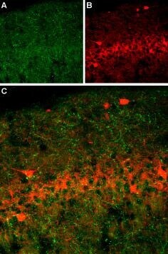
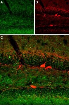
- Expression of SERT in rat brain - Immunohistochemical staining of immersion-fixed, free floating rat brain frozen sections using Anti-Serotonin Transporter (SERT) (extracellular) Antibody (#AMT-004), (1:400). A. SERT (green) was visualized in axonal fibers of the rat hippocampal CA1 region. B. Axonal fibers of neurons expressing gamma amino butyric acid (GABA) were labeled with mouse anti parvalbumin (red). C. Merge of the two images demonstrates separate axonal processes (no co-localization).
- Submitted by
- Invitrogen Antibodies (provider)
- Main image

- Experimental details
- Expression of SERT in mouse brain - Immunohistochemical staining of immersion-fixed, free floating mouse brain frozen sections using Anti-Serotonin Transporter (SERT) (extracellular) Antibody (#AMT-004), (1:400). A. SERT (green) was visualized in axonal fibers of the hippocampal dentate gyrus. B. Axonal fibers of neurons expressing gamma amino butyric acid (GABA) were labeled with mouse anti parvalbumin (red). C. Merge of the two images demonstrates separate axonal processes (no co-localization).
Supportive validation
- Submitted by
- Invitrogen Antibodies (provider)
- Main image

- Experimental details
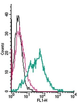
- Cell surface detection of SERT by indirect flow cytometry in live intact humanMEG-01 megakaryocyticleukemiacells: - (black line) cells. (red) Cells+ goat- Anti-rabbit-FITC. (green) Cells Anti-Serotonin Transporter (SERT) (extracellular) Antibody (#AMT-004), (2.5μg) + goat- Anti-rabbit-FITC.