Explore
 Validate
 Learn
Learn Western blot
Antibody data
- Antibody Data
- Antigen structure
- References [2]
- Comments [0]
- Validations
- Western blot [4]
- Immunocytochemistry [3]
- Immunoprecipitation [1]
- Flow cytometry [1]
Submit
Validation data
Reference
Comment
Report error
- Product number
- GTX100622 - Provider product page

- Provider
- GeneTex
- Proper citation
- GeneTex Cat#GTX100622, RRID:AB_2037563
- Product name
- Oct4 antibody
- Antibody type
- Polyclonal
- Reactivity
- Human, Mouse
- Host
- Rabbit
Submitted references An RNA-binding Protein, Lin28, Recognizes and Remodels G-quartets in the MicroRNAs (miRNAs) and mRNAs It Regulates.
Up-regulation of FOXM1 by E6 oncoprotein through the MZF1/NKX2-1 axis is required for human papillomavirus-associated tumorigenesis.
O'Day E, Le MTN, Imai S, Tan SM, Kirchner R, Arthanari H, Hofmann O, Wagner G, Lieberman J
The Journal of biological chemistry 2015 Jul 17;290(29):17909-17922
The Journal of biological chemistry 2015 Jul 17;290(29):17909-17922
Up-regulation of FOXM1 by E6 oncoprotein through the MZF1/NKX2-1 axis is required for human papillomavirus-associated tumorigenesis.
Chen PM, Cheng YW, Wang YC, Wu TC, Chen CY, Lee H
Neoplasia (New York, N.Y.) 2014 Nov;16(11):961-71
Neoplasia (New York, N.Y.) 2014 Nov;16(11):961-71
No comments: Submit comment
Supportive validation
- Submitted by
- GeneTex (provider)
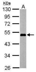
- Main image

- Experimental details
- Sample (20 ?g) A: HeLa nucleus 10% SDS PAGE GTX100622 diluted at 1:1000 The HRP-conjugated anti-rabbit IgG antibody (GTX213110-01) was used to detect the primary antibody.

- Submitted by
- GeneTex (provider)
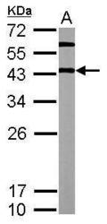
- Main image
- Experimental details
- Sample (20 ?g of whole cell lysate) A: Human ESC 10% SDS PAGE GTX100622 diluted at 1:1000 The HRP-conjugated anti-rabbit IgG antibody (GTX213110-01) was used to detect the primary antibody.

- Submitted by
- GeneTex (provider)
- Main image
- Experimental details
- Sample (20 ?g of whole cell lysate) A: mouse ESC 12% SDS PAGE GTX100622 diluted at 1:5000 The HRP-conjugated anti-rabbit IgG antibody (GTX213110-01) was used to detect the primary antibody.

- Submitted by
- GeneTex (provider)
- Main image
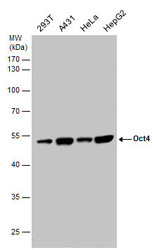
- Experimental details
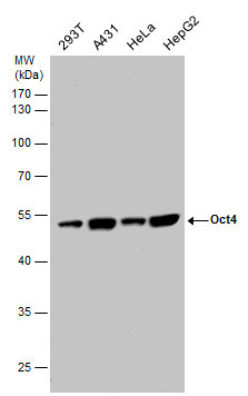
- OCT4 antibody detects OCT4 protein by western blot analysis. Various whole cell extracts (30 ?g) were separated by 10% SDS-PAGE, and the membrane was blotted with OCT4 antibody (GTX100622) diluted at a dilution of 1:10000. The HRP-conjugated anti-rabbit IgG antibody (GTX213110-01) was used to detect the primary antibody.
Supportive validation
- Submitted by
- GeneTex (provider)
- Main image

- Experimental details
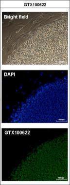
- Immunofluorescence analysis of paraformaldehyde-fixed human embryonic stem cell, using Oct4(GTX100622) antibody at 1:200 dilution.
- Submitted by
- GeneTex (provider)
- Main image
- Experimental details
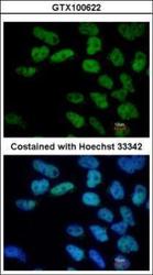
- Immunofluorescence analysis of paraformaldehyde-fixed Human ESC, using Oct4(GTX100622) antibody at 1:400 dilution.

- Submitted by
- GeneTex (provider)
- Main image
- Experimental details
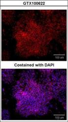
- Immunofluorescence analysis of paraformaldehyde-fixed human embryonic stem cell, using Oct4(GTX100622) antibody at 1:100 dilution.

Supportive validation
- Submitted by
- GeneTex (provider)
- Main image
- Experimental details
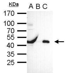
- Oct4 antibody immunoprecipitates Oct4 protein in IP experiments. IP Sample: cell lysate/extract of Oct4 gene transfected 293T cells A. Cell lysate/extract of transfected 293T cell B. Control with 2 £gg of preimmune rabbit IgG C. Immunoprecipitation of Oct4 by 2 £gg of Oct4 antibody (GTX100622) 12% SDS-PAGE The immunoprecipitated Oct4 protein was detected by Oct4 antibody (GTX100622) diluted at 1:1000. EasyBlot anti-rabbit IgG (GTX221666-01) was used as a secondary reagent.

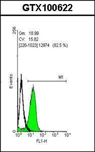
Supportive validation
- Submitted by
- GeneTex (provider)
- Main image

- Experimental details
- Flow cytometry on human embryonic stem cells, staining with Oct4 (GTX100622)antibody at 1:50 dilution(green) or rabbit IgG (black).