Explore
 Validate
 Learn
Learn Western blot
 Immunocytochemistry
Antibody data
- Antibody Data
- Antigen structure
- References [5]
- Comments [0]
- Validations
- Immunocytochemistry [1]
- Flow cytometry [1]
- Chromatin Immunoprecipitation [1]
- Other assay [1]
Submit
Validation data
Reference
Comment
Report error
- Product number
- 701756 - Provider product page

- Provider
- Invitrogen Antibodies
- Product name
- OCT4 Recombinant Rabbit Monoclonal Antibody (3H8L6)
- Antibody type
- Monoclonal
- Antigen
- Other
- Reactivity
- Human, Mouse
- Host
- Rabbit
- Isotype
- IgG
- Antibody clone number
- 3H8L6
- Vial size
- 100 µg
- Concentration
- 0.5 mg/mL
- Storage
- Store at 4°C short term. For long term storage, store at -20°C, avoiding freeze/thaw cycles.
Submitted references Effects of DMSO on the Pluripotency of Cultured Mouse Embryonic Stem Cells (mESCs).
Differentiation of Human Embryonic Stem Cells into Neuron, Cholinergic, and Glial Cells.
Four induced pluripotent stem cell lines (TRNDi021-C, TRNDi023-D, TRNDi024-D and TRNDi025-A) generated from fibroblasts of four healthy individuals.
Dysfunction of iPSC-derived endothelial cells in human Hutchinson-Gilford progeria syndrome.
Generation of two induced pluripotent stem cell lines (NHLBIi001-A and NHLBIi001-B) from a healthy Caucasian female volunteer with normal cardiac function.
Sousa MI, Correia B, Branco AF, Rodrigues AS, Ramalho-Santos J
Stem cells international 2020;2020:8835353
Stem cells international 2020;2020:8835353
Differentiation of Human Embryonic Stem Cells into Neuron, Cholinergic, and Glial Cells.
Hosseini K, Lekholm E, Ahemaiti A, Fredriksson R
Stem cells international 2020;2020:8827874
Stem cells international 2020;2020:8827874
Four induced pluripotent stem cell lines (TRNDi021-C, TRNDi023-D, TRNDi024-D and TRNDi025-A) generated from fibroblasts of four healthy individuals.
Xu X, Pradhan M, Xu M, Cheng YS, Beers J, Linask KL, Lin Y, Zheng W, Zou J
Stem cell research 2020 Dec;49:102011
Stem cell research 2020 Dec;49:102011
Dysfunction of iPSC-derived endothelial cells in human Hutchinson-Gilford progeria syndrome.
Matrone G, Thandavarayan RA, Walther BK, Meng S, Mojiri A, Cooke JP
Cell cycle (Georgetown, Tex.) 2019 Oct;18(19):2495-2508
Cell cycle (Georgetown, Tex.) 2019 Oct;18(19):2495-2508
Generation of two induced pluripotent stem cell lines (NHLBIi001-A and NHLBIi001-B) from a healthy Caucasian female volunteer with normal cardiac function.
Patterson K, Beers J, Linask KL, Lin Y, Hassanzadeh S, Sack MN, Zou J
Stem cell research 2019 Dec;41:101627
Stem cell research 2019 Dec;41:101627
No comments: Submit comment
Supportive validation
- Submitted by
- Invitrogen Antibodies (provider)
- Main image
- Experimental details
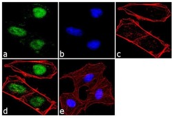
- For immunofluorescence analysis, NTERA-2 cells were fixed and permeabilized for detection of endogenous OCT4 using Anti- OCT4 Recombinant Rabbit Monoclonal Antibody (Product # 701756, 2 µg/mL) and labeled with Goat anti-Rabbit IgG (H+L) Superclonal™ Secondary Antibody, Alexa Fluor® 488 conjugate (Product # A27034, 1:2000). Panel a) shows representative cells that were stained for detection and localization of OCT4 protein (green), Panel b) is stained for nuclei (blue) using SlowFade® Gold Antifade Mountant with DAPI (Product # S36938). Panel c) represents cytoskeletal F-actin staining using Alexa Fluor® 555 Rhodamine Phalloidin (Product # R415, 1:300). Panel d) is a composite image of Panels a, b and c clearly demonstrating nuclear localization of OCT4. Panel e) represents control cells with no primary antibody to assess background. The images were captured at 60X magnification.

Supportive validation
- Submitted by
- Invitrogen Antibodies (provider)
- Main image
- Experimental details
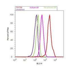
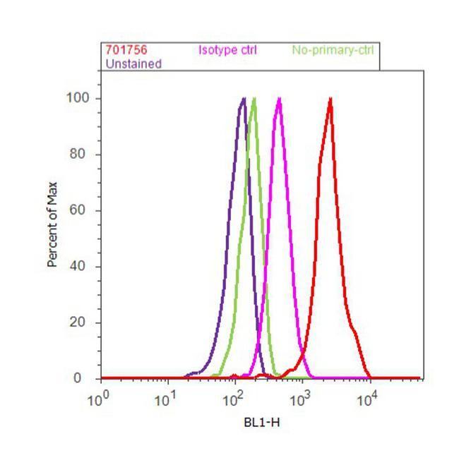
- Flow Cytometry analysis of endogenous OCT4 was performed on NTERA-2 cells labeled with ABfinity™ Anti-OCT4 Recombinant Rabbit Monoclonal Antibody (Product # 701756, 5 ug/1M cells) or with rabbit isotype control at 0.5 ug/ml and detected with Goat anti-Rabbit IgG (H+L) Superclonal™ Secondary Antibody, (Alexa Fluor® 488 conjugate, Product # A27034, 0.4 ug/ml, 1:2500) as represented by the red and pink histograms respectively. The purple histogram represents unstained control cells and the green histogram represents no-primary-antibody control. A representative of 10,000 cells were acquired and analyzed for each sample using an Attune® Acoustic Focusing Cytometer (4468770).
Supportive validation
- Submitted by
- Invitrogen Antibodies (provider)
- Main image
- Experimental details
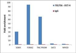
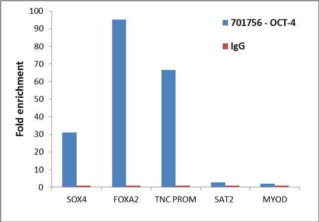
- Enrichment of endogenous OCT4 protein at specific gene loci using Anti-OCT4 Recombinant Rabbit Monoclonal Antibody: Chromatin Immunoprecipitation (ChIP) was performed using Anti-OCT4 Recombinant Rabbit Monoclonal Antibody (Product # 701756, 5 µg) on sheared chromatin from 2 million NTERA2 cells using the "MAGnify ChIP system" kit (Product # 49-2024). Normal Rabbit IgG (1 µg) was used as a negative IP control. The purified DNA was analyzed by 7500 Fast qPCR system (Product # 4351106) with optimized PCR primer pairs for the promoters of the active SOX4, FOXA2, TNC region used as positive control target genes, and the region of the inactive MYOD, SAT2 satellite repeat, used as negative control target gene. Data is presented as fold enrichment of the antibody signal versus the negative control IgG using the comparative CT method.
Supportive validation
- Submitted by
- Invitrogen Antibodies (provider)
- Main image
- Experimental details
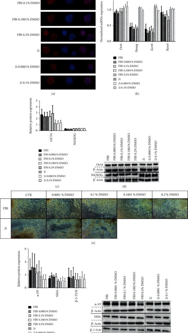
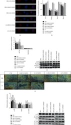
- Figure 3 Exposure to small percentages of DMSO for 48 hours does not negatively affect pluripotency marker expression or the differentiation potential of mESC. (a) Representative immunofluorescence images of plated colonies presenting the pluripotency marker Nanog (red) and counterstained with Hoechst 33342 (blue) (600x magnification) after a 48 h incubation with/without DMSO. (b) RT-PCR analysis for Oct4 , Nanog , Rexo1 , and Essrb mRNA gene expression normalized for endogenous beta-actin ( Actb ) at the 48 h time point. At least four independent experiments were performed, and the results are presented as means +- SEM. (c, d) Relative protein expression of the pluripotency factors Oct4 and Nanog evaluated by Western blot analysis and normalized by beta -actin expression at the 48 h time point. At least three independent experiments were performed, and the results are presented as means +- SEM. (e-g) Embryoid bodies (EBs) were generated from mESCs cultured for 48 h in the presence/absence of DMSO. After the differentiation protocol, every condition was able to generate fully differentiated cultures as shown in (e) by phase-contrast microscopy (100x magnification). (f, g) Western blot analysis and quantification of the protein levels of the three embryonic leaflet markers alpha -FP, SMA, and beta -3-TUB normalized by the expression of the loading control beta -actin. At least four independent experiments were performed, and the results are presented as means +- SEM. Statistic