 Explore
 Validate
 Learn
Learn Immunocytochemistry
Antibody data
- Antibody Data
- Antigen structure
- References [2]
- Comments [0]
- Validations
- Immunocytochemistry [1]
- Flow cytometry [2]
- Other assay [2]
Submit
Validation data
Reference
Comment
Report error
- Product number
- 53-5841-80 - Provider product page

- Provider
- Invitrogen Antibodies
- Product name
- Anti-OCT3/4 Monoclonal Antibody (EM92), Alexa Fluor 488, eBioscience™
- Antibody type
- Monoclonal
- Antigen
- Other
- Description
- Description: The EM92 monoclonal antibody reacts with mouse and human Oct3/4, encoded by the Pou5F1 gene. Oct3/4 is a POU domain-containing transcription factor that is critical for maintaining embryonic stem (ES) and induced pluripotent stem (iPS) cells in a pluripotent state, and is expressed by ES, embryonic germ cells and embryonic carcinoma cell lines. In cells of the inner cell mass (ICM), reduction of Oct3/4 expression causes dedifferentiation to trophoectoderm, whereas increased expression results in differentiation to mesoderm and primitive endoderm. Oct3/4 regulates the expression of several genes, including FGF-4, UTF1, Sox2, Fbx15, Rex1 and osteopontin through distinct mechanisms. Furthermore, Oct3/4 frequently acts synergistically with Sox2 to regulate target gene expression, as is the case with FGF-4. It has been demonstrated that Oct3/4 expression in ES cells can be negatively regulated by either treatment with retinoic acid, or by removal of leukemia-inhibitory factor (LIF). Applications Reported: This EM92 antibody has been reported for use in intracellular staining followed by flow cytometric analysis and immunocytochemistry. Applications Tested: This EM92 antibody has been tested by microscopy or intracellular staining and flow cytometric analysis of F9 cells using the Foxp3/Transcription Factor Staining Buffer Set (cat. 00-5523) and protocol. Please see Best Protocols for Staining Protocol (refer to Protocol B: One step protocol for intracellular (nuclear) proteins). This antibody can be used at less than or equal to 1 µg per test. A test is defined as the amount (µg) of antibody that will stain a cell sample in a final volume of 100 µL. Cell number should be determined empirically but can range from 10^5 to 10^8 cells/test. This antibody has also been tested by immunocytochemistry on formaldehyde fixed and permeabilized cells at less than or equal to 10µg/mL. It is recommended that the antibody be carefully titrated for optimal performance in the assay of interest. Excitation: 488 nm; Emission: 519 nm; Laser: Blue Laser. Filtration: 0.2 µm post-manufacturing filtered.
- Reactivity
- Human, Mouse
- Host
- Rat
- Conjugate
- Green dye
- Isotype
- IgG
- Antibody clone number
- EM92
- Vial size
- 25 µg
- Concentration
- 0.5 mg/mL
- Storage
- 4° C, store in dark, DO NOT FREEZE!
Submitted references Identification of a Hematopoietic Cell Dedifferentiation-Inducing Factor.
Highly upregulated Lhx2 in the Foxn1-/- nude mouse phenotype reflects a dysregulated and expanded epidermal stem cell niche.
Li Y, Adomat H, Guns ET, Hojabrpour P, Duronio V, Curran TA, Jalili RB, Jia W, Delwar Z, Zhang Y, Elizei SS, Ghahary A
Journal of cellular physiology 2016 Jun;231(6):1350-63
Journal of cellular physiology 2016 Jun;231(6):1350-63
Highly upregulated Lhx2 in the Foxn1-/- nude mouse phenotype reflects a dysregulated and expanded epidermal stem cell niche.
Bohr S, Patel SJ, Vasko R, Shen K, Huang G, Yarmush ML, Berthiaume F
PloS one 2013;8(5):e64223
PloS one 2013;8(5):e64223
No comments: Submit comment
Supportive validation
- Submitted by
- Invitrogen Antibodies (provider)
- Main image

- Experimental details
- Immunocytochemistry of human embryonic stem cells using 10 µg/mL Anti-OCT3/4 Alexa Fluor® 488 (Green) and 5 µg/mL Anti-Human TRA-1-81 Purified (Product # 53-5841-82) followed by Anti-Mouse TRITC (Red) (Right). Data courtesy of Zeng lab at the Buck Institute.
- Conjugate
- Green dye

Supportive validation
- Submitted by
- Invitrogen Antibodies (provider)
- Main image

- Experimental details
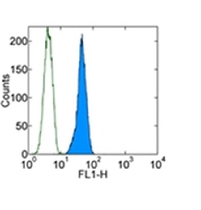
- Intracellular staining of F9 cell line with 0.5 µg of Rat IgG2a kappa Isotype Control Alexa Fluor® 488 (Product # 53-4321-80) (open histogram) or 0.5 µg of Anti-OCT3/4 Alexa Fluor® 488 (filled histogram) using Foxp3 Staining Buffers (Product # 00-5523-00). Total cells were used for analysis.
- Conjugate
- Green dye
- Submitted by
- Invitrogen Antibodies (provider)
- Main image
- Experimental details
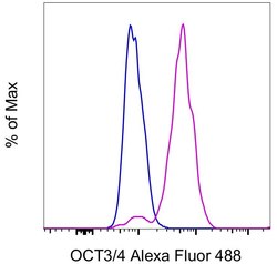
- F9 cells were stained intracellularly, using the Foxp3/Transcription Factor Staining Buffer Set (Product # 00-5523-00) and protocol, with 0.5 µg of Rat IgG2a kappa Isotype Control, Alexa Fluor 488 (Product # 53-4321-80) (blue histogram) or 0.5 µg of OCT3/4 Monoclonal Antibody, Alexa Fluor 488 (purple histogram). Total cells were used for analysis.
- Conjugate
- Green dye

Supportive validation
- Submitted by
- Invitrogen Antibodies (provider)
- Main image

- Experimental details
- Immunofluorescence analysis of OCT3/4 was performed using 70% confluent log phase NTERA-2 cells. The cells were fixed with 4% paraformaldehyde for 10 minutes, permeabilized with 0.1% Triton™ X-100 for 15 minutes, and blocked with 1% BSA for 1 hour at room temperature. The cells were labeled with OCT3/4 Mouse Monoclonal Antibody (Product # 53-5841-80) at 5ug/ml in 0.1% BSA, incubated at 4 degree Celsius overnight (Panel a: green). Nuclei (Panel b: blue) were stained with ProLong™ Diamond Antifade Mountant with DAPI (Product # P36962). F-actin (Panel c: red) was stained with Rhodamine Phalloidin (Product # R415, 1:300). Panel d represents the merged image showing nuclear localization. Panel e shows OCT3/4 negative cell line HeLa with no signal. Panel f represents control cells with Isotype control to assess background. The images were captured at 60X magnification.
- Conjugate
- Green dye

- Submitted by
- Invitrogen Antibodies (provider)
- Main image

- Experimental details
- Immunofluorescence analysis of OCT3/4 was performed using 70% confluent log phase NTERA cells. The cells were fixed with 4% paraformaldehyde for 10 minutes, permeabilized with 0.1% Triton™ X-100 for 15 minutes, and blocked with 1% BSA for 1 hour at room temperature. The cells were labeled with OCT3/4 Mouse Monoclonal Antibody (Product # 53-5841-82) at 5ug/ml in 0.1% BSA, incubated at 4 degree Celsius overnight (Panel a: green). Nuclei (Panel b: blue) were stained with ProLong™ Diamond Antifade Mountant with DAPI (Product # P36962). F-actin (Panel c: red) was stained with Rhodamine Phalloidin (Product # R415, 1:300). Panel d represents the merged image showing nuclear localization. Panel e shows OCT3/4 negative cell line HeLa with no signal. Panel f represents control cells with Isotype control to assess background. The images were captured at 60X magnification.
- Conjugate
- Green dye
