Explore
 Validate
 Learn
Learn Western blot
 Immunocytochemistry
Antibody data
- Antibody Data
- Antigen structure
- References [1]
- Comments [0]
- Validations
- Immunocytochemistry [6]
- Immunoprecipitation [2]
- Immunohistochemistry [5]
- Flow cytometry [2]
- Other assay [4]
Submit
Validation data
Reference
Comment
Report error
- Product number
- MA5-31458 - Provider product page

- Provider
- Invitrogen Antibodies
- Product name
- OCT4 Monoclonal Antibody (GT486)
- Antibody type
- Monoclonal
- Antigen
- Recombinant full-length protein
- Description
- Keep as concentrated solution. Predicted reactivity: Cat (89%), Pig (88%), Rabbit (85%), Rhesus Monkey (97%), Chimpanzee (100%), Bovine (85%). Positive Control: NT2D1, human ESC, mouse ESC, HeLa transfected recombinant OCT3/4 protein. Store product as a concentrated solution. Centrifuge briefly prior to opening the vial.
- Reactivity
- Human, Mouse
- Host
- Mouse
- Isotype
- IgG
- Antibody clone number
- GT486
- Vial size
- 100 μL
- Concentration
- 1 mg/mL
- Storage
- Store at 4°C short term. For long term storage, store at -20°C, avoiding freeze/thaw cycles.
Submitted references Hypoxia-induced autophagy drives colorectal cancer initiation and progression by activating the PRKC/PKC-EZR (ezrin) pathway.
Qureshi-Baig K, Kuhn D, Viry E, Pozdeev VI, Schmitz M, Rodriguez F, Ullmann P, Koncina E, Nurmik M, Frasquilho S, Nazarov PV, Zuegel N, Boulmont M, Karapetyan Y, Antunes L, Val D, Mittelbronn M, Janji B, Haan S, Letellier E
Autophagy 2020 Aug;16(8):1436-1452
Autophagy 2020 Aug;16(8):1436-1452
No comments: Submit comment
Supportive validation
- Submitted by
- Invitrogen Antibodies (provider)
- Main image
- Experimental details
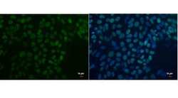
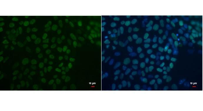
- Oct4 antibody detects Oct4 protein by immunofluorescent analysis. Sample: human embryonic stem cell were fixed in 4% paraformaldehyde for 15 min. Green: Oct4 protein stained by Oct4 antibody (Product # MA5-31458) diluted at 1:200. Blue: Hoechst 33342 staining. Scale bar = 10 µm.
- Submitted by
- Invitrogen Antibodies (provider)
- Main image

- Experimental details
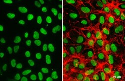
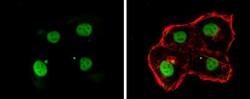
- Immunocytochemistry-Immunofluorescence analysis of OCT4 was performed in NT2D1 cells fixed in 4% paraformaldehyde at RT for 15 min. Green: OCT4 Monoclonal Antibody (GT486) (Product # MA5-31458) diluted at 1:500. Red: phalloidin, a cytoskeleton marker.

- Submitted by
- Invitrogen Antibodies (provider)
- Main image
- Experimental details
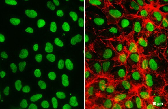
- OCT4 Monoclonal Antibody (GT486) detects Oct4 protein at nucleus by immunofluorescent analysis. Sample: NT2D1 cells were fixed in 4% paraformaldehyde at RT for 15 min. Green: Oct4 stained by OCT4 Monoclonal Antibody (GT486) (Product # MA5-31458) diluted at 1:500. Red: phalloidin, a cytoskeleton marker, diluted at 1:200. Scale bar= 10 µm.

- Submitted by
- Invitrogen Antibodies (provider)
- Main image

- Experimental details
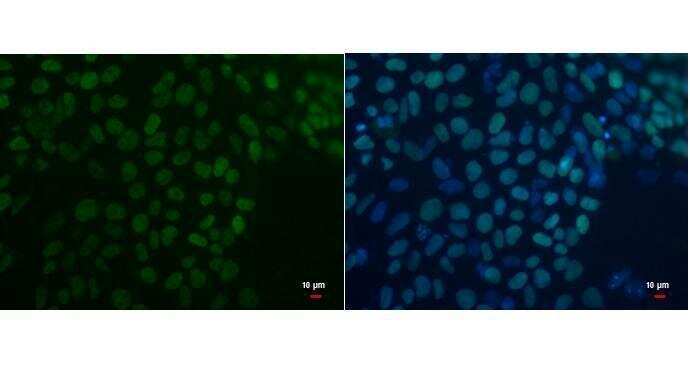
- Immunocytochemistry analysis of OCT4 in 4% paraformaldehyde-fixed human embryonic stem cells using OCT4 monoclonal antibody (Product # MA5-31458) at a dilution of 1:200. Sample was then incubated with Hoechst secondary antibody.
- Submitted by
- Invitrogen Antibodies (provider)
- Main image

- Experimental details
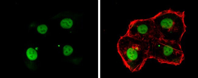
- OCT4 Monoclonal Antibody (GT486) detects Oct4 protein at nucleus by immunofluorescent analysis. Sample: NT2D1 cells were fixed in 4% paraformaldehyde at RT for 15 min. Green: Oct4 stained by OCT4 Monoclonal Antibody (GT486) (Product # MA5-31458) diluted at 1:500. Red: phalloidin, a cytoskeleton marker, diluted at 1:200. Scale bar= 10 µm.
- Submitted by
- Invitrogen Antibodies (provider)
- Main image
- Experimental details
- Immunocytochemistry-Immunofluorescence analysis of OCT4 was performed in NT2D1 cells fixed in 4% paraformaldehyde at RT for 15 min. Green: OCT4 Monoclonal Antibody (GT486) (Product # MA5-31458) diluted at 1:500. Red: phalloidin, a cytoskeleton marker.
Supportive validation
- Submitted by
- Invitrogen Antibodies (provider)
- Main image

- Experimental details
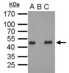
- Oct4 antibody immunoprecipitates Oct4 protein in IP experiments. IP Sample: cell lysate/extract of Oct4 gene transfected 293T cells A. Cell lysate/extract of transfected 293T cell B. Control with 2 µg of preimmune mouse IgG C. Immunoprecipitation of Oct4 by 2 µg of Oct4 antibody (Product # MA5-31458) 12% SDS-PAGE The immunoprecipitated Oct4 protein was detected by Oct4 antibody (Product # MA5-31458) diluted at 1:1,000.

- Submitted by
- Invitrogen Antibodies (provider)
- Main image

- Experimental details
- Immunoprecipitation analysis of OCT4 in transfected 293T cells, A) cell lysate of transfected 293T cell, B) control with preimmune mouse IgG, C) immunoprecipitation of Oct4 protein with OCT4 monoclonal antibody (Product # MA5-31458) using 2 µg of sample at a dilution of 1:1000. Sample was then incubated with anti-mouse IgG secondary antibody. Prior to incubation with primary antibody, the sample was separated on 12% SDS-PAGE.

Supportive validation
- Submitted by
- Invitrogen Antibodies (provider)
- Main image

- Experimental details
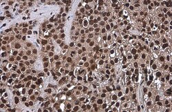
- Immunohistochemical analysis of paraffin-embedded BT483 xenograft, using OCT3/4 (Product # MA5-31458) antibody at 1:200 dilution. Antigen Retrieval: EDTA based buffer, pH 8.0, 15 min.

- Submitted by
- Invitrogen Antibodies (provider)
- Main image
- Experimental details
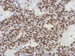
- Immunohistochemistry (Paraffin) analysis of OCT4 was performed in paraffin-embedded human cervical carcinoma tissue using OCT4 Monoclonal Antibody (GT486) (Product # MA5-31458) at a dilution of 1:200. Antigen Retrieval: Citrate buffer, pH 6.0, 15 min.
- Submitted by
- Invitrogen Antibodies (provider)
- Main image
- Experimental details
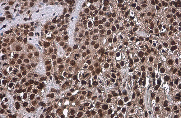
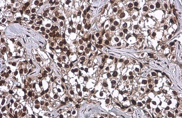
- OCT4 Monoclonal Antibody (GT486) detects Oct4 protein at nucleus by immunohistochemical analysis. Sample: Paraffin-embedded human breast carcinoma. Oct4 stained by OCT4 Monoclonal Antibody (GT486) (Product # MA5-31458) diluted at 1:500. Antigen Retrieval: Citrate buffer, pH 6.0, 15 min.
- Submitted by
- Invitrogen Antibodies (provider)
- Main image
- Experimental details
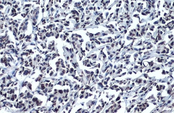
- Immunohistochemistry (Paraffin) analysis of OCT4 was performed in paraffin-embedded human breast carcinoma tissue using OCT4 Monoclonal Antibody (GT486) (Product # MA5-31458) at a dilution of 1:200. Antigen Retrieval: Citrate buffer, pH 6.0, 15 min.
- Submitted by
- Invitrogen Antibodies (provider)
- Main image
- Experimental details
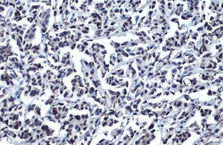
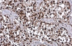
- Immunohistochemistry analysis of OCT4 in paraffin-embedded BT483 xenograft using OCT4 monoclonal antibody (Product # MA5-31458) at a dilution of 1:200.

Supportive validation
- Submitted by
- Invitrogen Antibodies (provider)
- Main image
- Experimental details
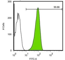
- OCT4 Monoclonal Antibody (GT486) (Product # MA5-31458) detects POU5F protein by flow cytometry analysis. Sample: Human embryonic stem cells Black: Isotype control dilution: 1:50 Green: OCT4 Monoclonal Antibody (GT486) (Product # MA5-31458) dilution: 1:50.

- Submitted by
- Invitrogen Antibodies (provider)
- Main image

- Experimental details
- Flow cytometry analysis of OCT4 in human embryonic stem cells using OCT4 monoclonal antibody (Product # MA5-31458) at a dilution of 1:50.

Supportive validation
- Submitted by
- Invitrogen Antibodies (provider)
- Main image
- Experimental details
- Immunoprecipitation analysis of OCT4 in transfected 293T cells, A) cell lysate of transfected 293T cell, B) control with preimmune mouse IgG, C) immunoprecipitation of Oct4 protein with OCT4 monoclonal antibody (Product # MA5-31458) using 2 µg of sample at a dilution of 1:1000. Sample was then incubated with anti-mouse IgG secondary antibody. Prior to incubation with primary antibody, the sample was separated on 12% SDS-PAGE.

- Submitted by
- Invitrogen Antibodies (provider)
- Main image
- Experimental details
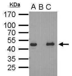
- Oct4 antibody immunoprecipitates Oct4 protein in IP experiments. IP Sample: cell lysate/extract of Oct4 gene transfected 293T cells A. Cell lysate/extract of transfected 293T cell B. Control with 2 µg of preimmune mouse IgG C. Immunoprecipitation of Oct4 by 2 µg of Oct4 antibody (Product # MA5-31458) 12% SDS-PAGE The immunoprecipitated Oct4 protein was detected by Oct4 antibody (Product # MA5-31458) diluted at 1:1,000.

- Submitted by
- Invitrogen Antibodies (provider)
- Main image

- Experimental details
- Oct4 antibody immunoprecipitates Oct4 protein in IP experiments. IP Sample: cell lysate/extract of Oct4 gene transfected 293T cells A. Cell lysate/extract of transfected 293T cell B. Control with 2 µg of preimmune mouse IgG C. Immunoprecipitation of Oct4 by 2 µg of Oct4 antibody (Product # MA5-31458) 12% SDS-PAGE The immunoprecipitated Oct4 protein was detected by Oct4 antibody (Product # MA5-31458) diluted at 1:1,000.

- Submitted by
- Invitrogen Antibodies (provider)
- Main image
- Experimental details
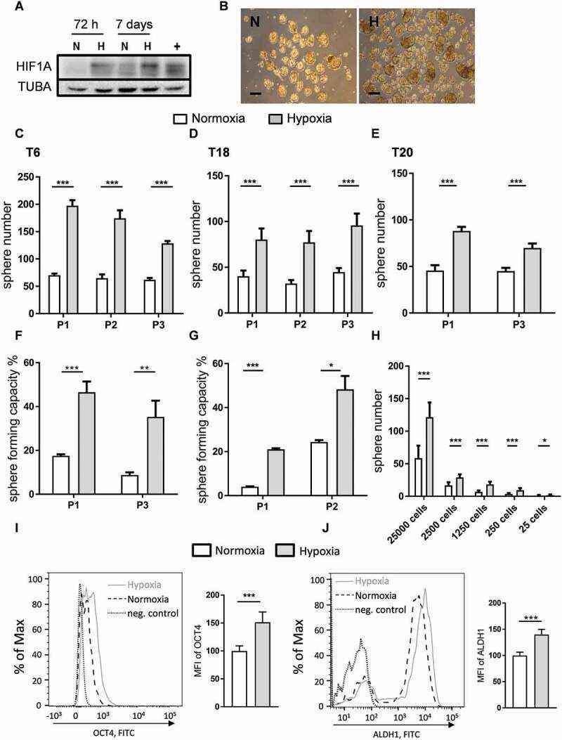
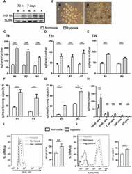
- Figure 1. Hypoxia increases the self-renewal capacity of patient-derived TIC cultures. ( A ) HIF1A protein expression under normoxic (N) and hypoxic (H) culturing conditions over 3 d (72 h) and 7 d in T6 TICs. TUBA staining was used as a loading control and HepG2 cells exposed to hypoxia for 24 h were used as a positive control (+). ( B ) Representative image of patient T6-derived TICs, cultured under normoxia (N) and hypoxia (H). Scale: 100 um. ( C-H ) Self-renewal capacity as determined by the 1000 cell sphere formation assay ( C-E ) in T6 ( C ), T18 ( D ) and T20 ( E ) cultures and by the single cell assay ( F-G ) in T6 ( F ) and T18 ( G ) cultures. Sphere formation was observed over several passages (P). ( H ) Self-renewal capacity was determined by a limiting dilution assay at multiple cell doses under normoxia and hypoxia. Results from one experiment using T6 TICs are shown and ELDA was used to assess significance. C-G; Data are representative of at least three independent experiments. ( I-J ) Hypoxia induces the expression of stem cell markers. ( I ) Flow cytometry staining of POU5F1 in T18 TICs under normoxic and hypoxic conditions and quantification of mean fluorescence intensity (MFI) of POU5F1 in two independent experiments (data normalized to normoxia). ( J ) Flow cytometry staining of ALDH1A1 in T18 TICs under normoxic and hypoxic conditions and quantification of mean fluorescence intensity (MFI) of ALDH1A1 in two independent experiments (data normalized to normo