 Explore
 Validate
 Learn
Learn Western blot
Antibody data
- Antibody Data
- Antigen structure
- References [4]
- Comments [0]
- Validations
- Western blot [4]
- Immunocytochemistry [1]
- Immunohistochemistry [2]
- Flow cytometry [4]
- Other assay [2]
Submit
Validation data
Reference
Comment
Report error
- Product number
- PA1-16943 - Provider product page

- Provider
- Invitrogen Antibodies
- Product name
- OCT4 Polyclonal Antibody
- Antibody type
- Polyclonal
- Antigen
- Synthetic peptide
- Description
- This antibody is expected to react with swine, feline and bovine samples based on a 93% sequence homology.
- Reactivity
- Human, Mouse, Rat, Bovine
- Host
- Rabbit
- Isotype
- IgG
- Vial size
- 100 µL
- Concentration
- 1 mg/mL
- Storage
- -20° C, Avoid Freeze/Thaw Cycles
Submitted references Characterization of mesenchymal stem cells derived from adipose tissue of a cougar (Puma concolor).
Whether CD44 is an applicable marker for glioma stem cells.
FXR-Gankyrin axis is involved in development of pediatric liver cancer.
The endometrium of cycling cows contains populations of putative mesenchymal progenitor cells.
Echeverry DM, Asenjo PA, Rojas DM, Aguilera CJ, Rodríguez-Álvarez L, Castro FO
Animal reproduction 2020 May 5;17(2):e20190109
Animal reproduction 2020 May 5;17(2):e20190109
Whether CD44 is an applicable marker for glioma stem cells.
Wang HH, Liao CC, Chow NH, Huang LL, Chuang JI, Wei KC, Shin JW
American journal of translational research 2017;9(11):4785-4806
American journal of translational research 2017;9(11):4785-4806
FXR-Gankyrin axis is involved in development of pediatric liver cancer.
Valanejad L, Lewis K, Wright M, Jiang Y, D'Souza A, Karns R, Sheridan R, Gupta A, Bove K, Witte D, Geller J, Tiao G, Nelson DL, Timchenko L, Timchenko N
Carcinogenesis 2017 Jul 1;38(7):738-747
Carcinogenesis 2017 Jul 1;38(7):738-747
The endometrium of cycling cows contains populations of putative mesenchymal progenitor cells.
Cabezas J, Lara E, Pacha P, Rojas D, Veraguas D, Saravia F, Rodríguez-Alvarez L, Castro FO
Reproduction in domestic animals = Zuchthygiene 2014 Aug;49(4):550-559
Reproduction in domestic animals = Zuchthygiene 2014 Aug;49(4):550-559
No comments: Submit comment
Supportive validation
- Submitted by
- Invitrogen Antibodies (provider)
- Main image

- Experimental details
- Western Blot detection of OCT4 in mouse brain lysate using Product # PA1-16943.

- Submitted by
- Invitrogen Antibodies (provider)
- Main image
- Experimental details
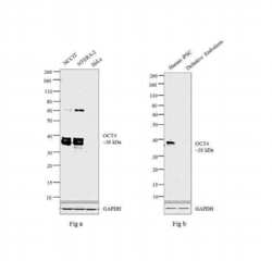
- Western blot analysis was performed on modified whole cell extracts (1% SDS) (30 µg lysate) of (Fig. a ) NCCIT (Lane 1), NTERA-2 (Lane 2) and HeLa (Lane 3), (Fig. b) Human iPSCs (Lane 1) and Human iPSCs derived definitive endoderm (Lane 2). The blot was probed with Anti- OCT4 Polyclonal Antibody (Product # PA1-16943, 1:1000 dilution) and detected by chemiluminescence using Goat anti-Rabbit IgG (H+L) Superclonal™ Secondary Antibody, HRP conjugate (Product # A27036, 0.25 µg/ml, 1:4000 dilution). A 38 kDa band corresponding to OCT4 was observed across the cell lines tested except for HeLa (Fig. a) and human iPSC derived definitive endoderm (Fig. b) which is reported to be negative for OCT4 expression.

- Submitted by
- Invitrogen Antibodies (provider)
- Main image
- Experimental details
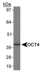
- Western blot analysis of OCT4 in mouse brain lysate. Samples were incubated in OCT4 polyclonal antibody (Product # PA1-16943). The antibody generated a specific band of OCT4 at the expected ~40 kDa position.

- Submitted by
- Invitrogen Antibodies (provider)
- Main image
- Experimental details
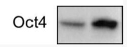
- Western blot analysis of OCT4 in human head and neck cancer cell lines. Sample was incubated in OCT4 polyclonal antibody (Product # PA1-16943).

Supportive validation
- Submitted by
- Invitrogen Antibodies (provider)
- Main image

- Experimental details
- Immunocytochemistry analysis of OCT4 in Ntera2 cells fixed in 4% paraformaldehyde for 10 minutes and permeabilized in 0.5% Triton X-100 in PBS for 5 minutes. Samples were incubated in OCT4 polyclonal antibody (Product # PA1-16943) using a dilution of 1 µg/mL overnight at 4 °C followed by anti-rabbit DyLight 488 (Green) with a dilution of 1:1000 dilution for 60 minutes. Nuclei were counterstained with DAPI (Blue). Cells were imaged using a 100X objective and digitally deconvolved.

Supportive validation
- Submitted by
- Invitrogen Antibodies (provider)
- Main image

- Experimental details
- OCT4 staining in NTERA-2 cells detected with a Dylight 488 labeled secondary antibody.

- Submitted by
- Invitrogen Antibodies (provider)
- Main image

- Experimental details
- Immunohistochemical analysis of OCT4 in formalin-fixed paraffin-embedded tissue section of mouse brain. Samples were incubated in OCT4 polyclonal antibody (Product # PA1-16943) using a dilution of 1:200 followed by HRP labeled anti-rabbit secondary antibody and DAB reagent. Nuclei of cells were counter-stained with hematoxylin.

Supportive validation
- Submitted by
- Invitrogen Antibodies (provider)
- Main image
- Experimental details
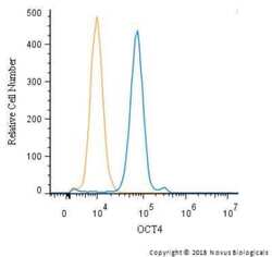
- FACS staining of NTERA-2 cells using Product # PA1-16943 at a 1:50 dilution detected using Dylight-488 conjugated goat anti-rabbit secondary antibody.

- Submitted by
- Invitrogen Antibodies (provider)
- Main image
- Experimental details
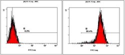
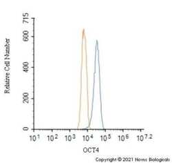
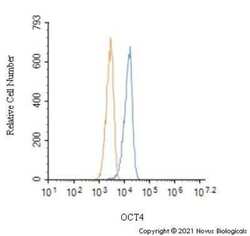
- Flow cytometry of OCT4 in Neuro2a cells (blue) and a matched isotype control (orange). Samples were incubated in OCT4 polyclonal antibody (Product # PA1-16943) using a dilution of 1.0 µg/mL for 30 minutes at room temperature followed by a Rabbit IgG (H+L) Cross-Adsorbed Secondary Antibody, Dylight™ 550 (Product # SA5-10033). Cells were fixed with 4% PFA and then permeabilized with 0.1% saponin.

- Submitted by
- Invitrogen Antibodies (provider)
- Main image
- Experimental details
- Flow cytometry of OCT4 in Jurkat cells (blue) and a matched isotype control (orange). Samples were incubated in OCT4 polyclonal antibody (Product # PA1-16943) using a dilution of 1.0 µg/mL for 30 minutes at room temperature followed by a Rabbit IgG (H+L) Cross-Adsorbed Secondary Antibody, Dylight™ 550 (Product # SA5-10033). Cells were fixed with 4% PFA and then permeabilized with 0.1% saponin.

- Submitted by
- Invitrogen Antibodies (provider)
- Main image
- Experimental details
- Flow cytometry of OCT4 in Jurkat and a matched isotype control. Samples were incubated in OCT4 polyclonal antibody (Product # PA1-16943) using a dilution of 1 µg/mL for 30 minutes at room temperature followed by a Rabbit IgG (H+L) Cross-Adsorbed secondary antibody. Cells were fixed with 4% PFA and then permeabilized with 0.1% saponin.

Supportive validation
- Submitted by
- Invitrogen Antibodies (provider)
- Main image

- Experimental details
- NULL

- Submitted by
- Invitrogen Antibodies (provider)
- Main image

- Experimental details
- Figure 4 Detection of OCT4 and SOX2 by immunocytochemistry in AMSC from Puma concolor . (a and c) negative controls of expression for OCT4 and SOX2 respectively; the cells were fixed, primary antibody was omitted and the cells were incubated with the secondary anti-rabbit HRP sheep conjugated antibody and revealed with DAB; (b) Strong detection of OCT4 in cytoplasm and nuclei and (d) SOX2 immunodetection in the cytoplasm of some cells. Antibodies, catalogue numbers and specificity are described in materials and methods.
