 Explore
 Validate
 Learn
Learn Western blot
 Immunocytochemistry
Antibody data
- Antibody Data
- Antigen structure
- References [9]
- Comments [0]
- Validations
- Immunocytochemistry [5]
- Immunoprecipitation [1]
- Immunohistochemistry [1]
- Flow cytometry [2]
- Other assay [7]
Submit
Validation data
Reference
Comment
Report error
- Product number
- PA5-27438 - Provider product page

- Provider
- Invitrogen Antibodies
- Product name
- OCT4 Polyclonal Antibody
- Antibody type
- Polyclonal
- Antigen
- Recombinant full-length protein
- Description
- Recommended positive controls: human ESC, mouse ESC, NT2D1. Predicted reactivity: Mouse (83%), Rat (83%), Cat (94%), Pig (93%), Rabbit (90%), Rhesus Monkey (98%), Chimpanzee (100%), Bovine (91%). Store product as a concentrated solution. Centrifuge briefly prior to opening the vial.
- Reactivity
- Human, Mouse
- Host
- Rabbit
- Isotype
- IgG
- Vial size
- 100 μL
- Concentration
- 1.37 mg/mL
- Storage
- Store at 4°C short term. For long term storage, store at -20°C, avoiding freeze/thaw cycles.
Submitted references Characterization of N-terminal RYR2 variants outside CPVT1 hotspot regions using patient iPSCs reveal pathogenesis and therapeutic potential.
Patient-specific, re-engineered cardiomyocyte model confirms the circumstance-dependent arrhythmia risk associated with the African-specific common SCN5A polymorphism p.S1103Y: Implications for the increased sudden deaths observed in black individuals during the COVID-19 pandemic.
Suppression of premature transcription termination leads to reduced mRNA isoform diversity and neurodegeneration.
Bone marrow-derived mesenchymal stem cells combined with gonadotropin therapy restore postnatal oogenesis of chemo-ablated ovaries in rats via enhancing very small embryonic-like stem cells.
In situ differentiation of human-induced pluripotent stem cells into functional cardiomyocytes on a coaxial PCL-gelatin nanofibrous scaffold.
Meagre Argyrosomus regius (Asso, 1801) Stem Spermatogonia: Histological Characterization, Immunostaining, In Vitro Proliferation, and Cryopreservation.
The master transcription factor SOX2, mutated in anophthalmia/microphthalmia, is post-transcriptionally regulated by the conserved RNA-binding protein RBM24 in vertebrate eye development.
Upregulation of stem cell markers ALDH1A1 and OCT4 as potential biomarkers for the early detection of cervical carcinoma.
The TDH-GCN5L1-Fbxo15-KBP axis limits mitochondrial biogenesis in mouse embryonic stem cells.
Stutzman MJ, Kim CSJ, Tester DJ, Hamrick SK, Dotzler SM, Giudicessi JR, Miotto MC, Gc JB, Frank J, Marks AR, Ackerman MJ
Stem cell reports 2022 Sep 13;17(9):2023-2036
Stem cell reports 2022 Sep 13;17(9):2023-2036
Patient-specific, re-engineered cardiomyocyte model confirms the circumstance-dependent arrhythmia risk associated with the African-specific common SCN5A polymorphism p.S1103Y: Implications for the increased sudden deaths observed in black individuals during the COVID-19 pandemic.
Hamrick SK, John Kim CS, Tester DJ, Giudicessi JR, Ackerman MJ
Heart rhythm 2022 May;19(5):822-827
Heart rhythm 2022 May;19(5):822-827
Suppression of premature transcription termination leads to reduced mRNA isoform diversity and neurodegeneration.
LaForce GR, Farr JS, Liu J, Akesson C, Gumus E, Pinkard O, Miranda HC, Johnson K, Sweet TJ, Ji P, Lin A, Coller J, Philippidou P, Wagner EJ, Schaffer AE
Neuron 2022 Apr 20;110(8):1340-1357.e7
Neuron 2022 Apr 20;110(8):1340-1357.e7
Bone marrow-derived mesenchymal stem cells combined with gonadotropin therapy restore postnatal oogenesis of chemo-ablated ovaries in rats via enhancing very small embryonic-like stem cells.
Ebrahim N, Al Saihati HA, Shaman A, Dessouky AA, Farid AS, Hussien NI, Mostafa O, Seleem Y, Sabry D, Saad AS, Emam HT, Hassouna A, Badr OAM, Saffaf BA, Forsyth NR, Salim RF
Stem cell research & therapy 2021 Sep 27;12(1):517
Stem cell research & therapy 2021 Sep 27;12(1):517
In situ differentiation of human-induced pluripotent stem cells into functional cardiomyocytes on a coaxial PCL-gelatin nanofibrous scaffold.
Sridharan D, Palaniappan A, Blackstone BN, Dougherty JA, Kumar N, Seshagiri PB, Sayed N, Powell HM, Khan M
Materials science & engineering. C, Materials for biological applications 2021 Jan;118:111354
Materials science & engineering. C, Materials for biological applications 2021 Jan;118:111354
Meagre Argyrosomus regius (Asso, 1801) Stem Spermatogonia: Histological Characterization, Immunostaining, In Vitro Proliferation, and Cryopreservation.
Zupa R, Martino NA, Marzano G, Dell'Aquila ME, Corriero A
Animals : an open access journal from MDPI 2020 May 14;10(5)
Animals : an open access journal from MDPI 2020 May 14;10(5)
The master transcription factor SOX2, mutated in anophthalmia/microphthalmia, is post-transcriptionally regulated by the conserved RNA-binding protein RBM24 in vertebrate eye development.
Dash S, Brastrom LK, Patel SD, Scott CA, Slusarski DC, Lachke SA
Human molecular genetics 2020 Mar 13;29(4):591-604
Human molecular genetics 2020 Mar 13;29(4):591-604
Upregulation of stem cell markers ALDH1A1 and OCT4 as potential biomarkers for the early detection of cervical carcinoma.
Tulake W, Yuemaier R, Sheng L, Ru M, Lidifu D, Abudula A
Oncology letters 2018 Nov;16(5):5525-5534
Oncology letters 2018 Nov;16(5):5525-5534
The TDH-GCN5L1-Fbxo15-KBP axis limits mitochondrial biogenesis in mouse embryonic stem cells.
Donato V, Bonora M, Simoneschi D, Sartini D, Kudo Y, Saraf A, Florens L, Washburn MP, Stadtfeld M, Pinton P, Pagano M
Nature cell biology 2017 Apr;19(4):341-351
Nature cell biology 2017 Apr;19(4):341-351
No comments: Submit comment
Supportive validation
- Submitted by
- Invitrogen Antibodies (provider)
- Main image
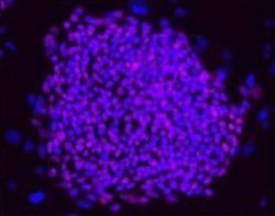
- Experimental details
- Immunofluorescent analysis of OCT3, 4 in hESCs on CF1 feeders cells using an OCT3, 4 polyclonal antibody (Product # PA5-27438) at a 1:200 dilution.
- Submitted by
- Invitrogen Antibodies (provider)
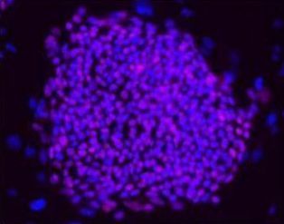
- Main image
- Experimental details
- Immunofluorescent analysis of OCT3, 4 in mESCs on CF1 feeders cells using an OCT3, 4 polyclonal antibody (Product # PA5-27438) at a 1:200 dilution.

- Submitted by
- Invitrogen Antibodies (provider)
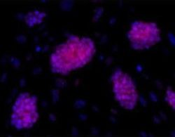
- Main image
- Experimental details
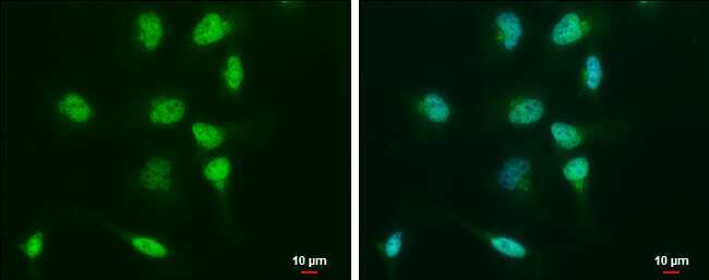
- Immunocytochemistry-Immunofluorescence analysis of OCT4 was performed in NT2D1 cells fixed in 4% paraformaldehyde at RT for 15 min. Green: OCT4 Polyclonal Antibody (Product # PA5-27438) diluted at 1:500. Blue: Hoechst 33342 staining. Scale bar = 10 µm.
- Submitted by
- Invitrogen Antibodies (provider)
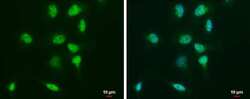
- Main image

- Experimental details
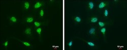
- Immunocytochemistry-Immunofluorescence analysis of OCT4 was performed in NT2D1 cells fixed in 4% paraformaldehyde at RT for 15 min. Green: OCT4 Polyclonal Antibody (Product # PA5-27438) diluted at 1:500. Blue: Hoechst 33342 staining. Scale bar = 10 µm.

- Submitted by
- Invitrogen Antibodies (provider)
- Main image
- Experimental details
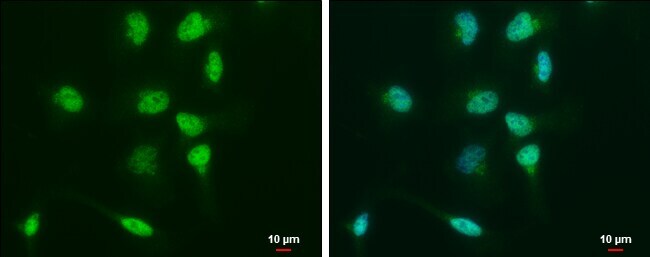
- Immunocytochemistry-Immunofluorescence analysis of OCT4 was performed in NT2D1 cells fixed in 4% paraformaldehyde at RT for 15 min. Green: OCT4 Polyclonal Antibody (Product # PA5-27438) diluted at 1:500. Blue: Hoechst 33342 staining. Scale bar = 10 µm.
Supportive validation
- Submitted by
- Invitrogen Antibodies (provider)
- Main image

- Experimental details
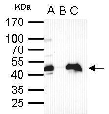
- OCT4 Polyclonal Antibody immunoprecipitates Oct4 protein in IP experiments. IP Sample: cell lysate/extract of Oct4 gene transfected 293T cells A. Cell lysate/extract of transfected 293T cell B. Control with 2 µg of preimmune rabbit IgG C. Immunoprecipitation of Oct4 by 2 µg of OCT4 Polyclonal Antibody (Product # PA5-27438) 12% SDS-PAGE The immunoprecipitated Oct4 protein was detected by OCT4 Polyclonal Antibody (Product # PA5-27438) diluted at 1:1,000.

Supportive validation
- Submitted by
- Invitrogen Antibodies (provider)
- Main image
- Experimental details
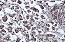
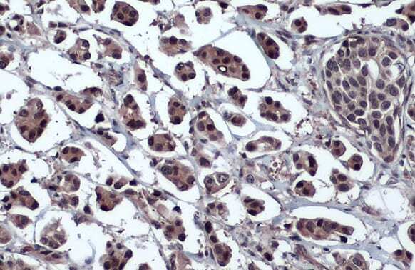
- Immunohistochemistry (Paraffin) analysis of OCT4 was performed in paraffin-embedded human breast carcinoma tissue using OCT4 Polyclonal Antibody (Product # PA5-27438) at a dilution of 1:1000. Antigen Retrieval: Citrate buffer, pH 6.0, 15 min.
Supportive validation
- Submitted by
- Invitrogen Antibodies (provider)
- Main image

- Experimental details
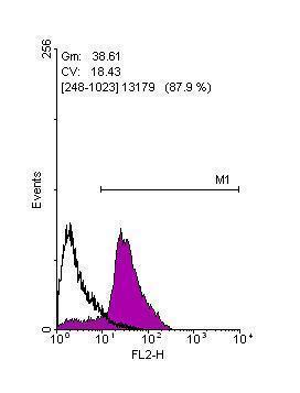
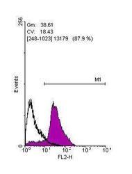
- Flow cytometry on human embryonic stem cells, staining with OCT4 Polyclonal Antibody (Product # PA5-27438) at 1:100 dilution (purple) or rabbit IgG (black).
- Submitted by
- Invitrogen Antibodies (provider)
- Main image
- Experimental details
- Flow cytometry on human embryonic stem cells, staining with OCT4 Polyclonal Antibody (Product # PA5-27438) at 1:100 dilution (purple) or rabbit IgG (black).

Supportive validation
- Submitted by
- Invitrogen Antibodies (provider)
- Main image
- Experimental details
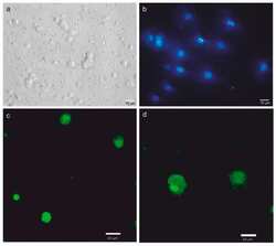
- Figure 6 Characterization of meagre spermatogonial stem cells (SSCs) cultured in vitro. Photomicrographs of cells exhibiting round-shape morphology, as observed via phase contrast microscopy ( a ), and presence of nuclei and small cytoplasm, as observed via epifluorescence microscopy after Hoechst staining ( b ). Confocal microscopy evaluation revealed anti-oct4- ( c ) and anti-vasa-positive ( d ) cells after 14 days of in vitro culture.

- Submitted by
- Invitrogen Antibodies (provider)
- Main image
- Experimental details
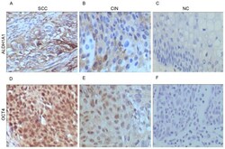
- Figure 2. Expression pattern of ALDH1 and OCT4 proteins in cervical lesions as detected by immunohistochemical staining. Expression of ALDH1A1 in (A) SCC, (B) CIN and (C) NC tissues. Expression of OCT4 in (D) SCC, (E) CIN and (F) NC tissues. Magnification, x400. ALDH1A1, aldehyde dehydrogenase 1 family member A1; OCT4, POU class 5 homeobox 1; SCC, cervical squamous cell carcinoma; CIN, cervical intraepithelial neoplasia; NC, negative controls.

- Submitted by
- Invitrogen Antibodies (provider)
- Main image

- Experimental details
- OCT4 Polyclonal Antibody immunoprecipitates Oct4 protein in IP experiments. IP Sample: cell lysate/extract of Oct4 gene transfected 293T cells A. Cell lysate/extract of transfected 293T cell B. Control with 2 µg of preimmune rabbit IgG C. Immunoprecipitation of Oct4 by 2 µg of OCT4 Polyclonal Antibody (Product # PA5-27438) 12% SDS-PAGE The immunoprecipitated Oct4 protein was detected by OCT4 Polyclonal Antibody (Product # PA5-27438) diluted at 1:1,000.
- Submitted by
- Invitrogen Antibodies (provider)
- Main image
- Experimental details
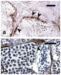
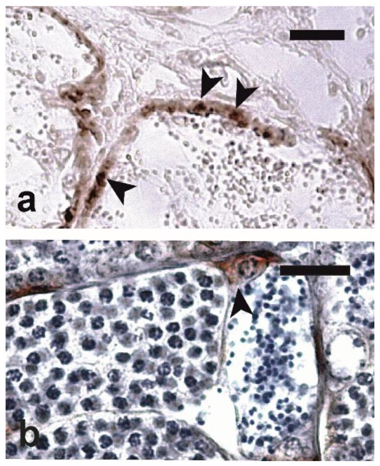
- Figure 2 Micrographs of testis sections of meagre immunostained with ( a ) anti-oct4 antibodies (Trial 1) and ( b ) anti-vasa antibodies (Trial 2). Intralobular positive cells (arrowheads) are stained in brown. In ( b ), Mayer's haematoxylin was used as a nuclear counterstain. Magnification bars = 20 um.
- Submitted by
- Invitrogen Antibodies (provider)
- Main image

- Experimental details
- Figure 6 Characterization of meagre spermatogonial stem cells (SSCs) cultured in vitro. Photomicrographs of cells exhibiting round-shape morphology, as observed via phase contrast microscopy ( a ), and presence of nuclei and small cytoplasm, as observed via epifluorescence microscopy after Hoechst staining ( b ). Confocal microscopy evaluation revealed anti-oct4- ( c ) and anti-vasa-positive ( d ) cells after 14 days of in vitro culture.

- Submitted by
- Invitrogen Antibodies (provider)
- Main image
- Experimental details
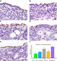
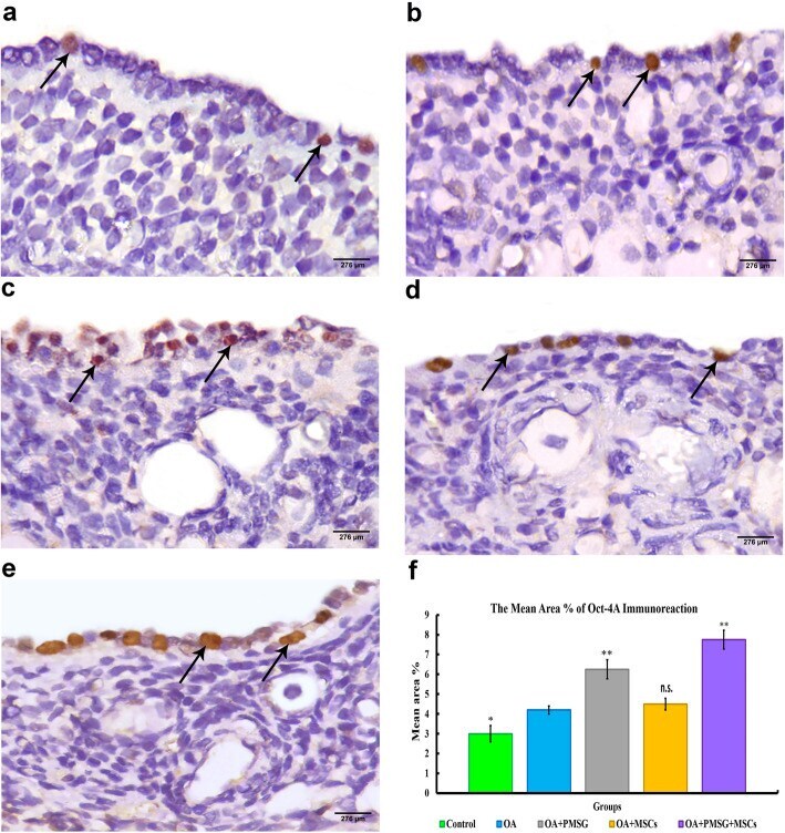
- Fig. 9 Representative photomicrograph of OCT4-A immunoreaction sections of different experimental groups. a Group I (control group) displayed minimal nuclear immuno-expression of OCT4-A in the surface epithelial cells. b Group II (chemo-ablated, OA group) showed mild nuclear OCT4-A immuno-expression in the surface epithelial cells. c Group III (OA + PMSG group) showed moderate nuclear OCT4A immuno-expression in the surface epithelial cells. d Group IV (OA + BM-MSCs group) showed minimal OCT4-A immuno-expression. e Group V (OA + PMSG+ BM-MSCs group) showed extensive immuno-expression of nuclear OCT4-A in the surface epithelial cells. f Histogram representing the mean area percentage of OCT4-A immunoreaction in all experimental groups. The significant differences against the chemo-ablated group indicated by a ""*"" for p < 0.05 and a ""**"" for p < 0.01. Data are shown as mean +- S.E.M, n = 7
- Submitted by
- Invitrogen Antibodies (provider)
- Main image
- Experimental details
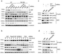
- Fig. 2 GCN5L1 and TDH are necessary for the Fbxo15-mediated degradation of KBP in mESCs a. mESCs were transfected with either a nontargeting (N/T) siRNA or siRNAs to Fbxo15 (oligo #1), GCN5L1, TDH, or KBP. Cells were either maintained in LIF-containing medium or induced to differentiate for 24 hours (24h) after LIF withdrawal and exposure to retinoic acid (RA). Cells were then collected and lysed for immunoblotting as indicated. The asterisk denotes an unspecific band. b. mESCs were transfected with a nontargeting (N/T) siRNA or siRNAs to Fbxo15 (oligo #2), GCN5L1, or TDH and treated with cycloheximide (CHX) for the indicated times. Cells were then collected and lysed for immunoblotting as indicated. c. mESCs were infected with lentiviruses expressing either HA-tagged wild type mouse KBP or HA-tagged mouse KBP(KK/RR) and then transfected with the indicated siRNAs. Whole cell extracts (WCE) were immunoprecipitated (IP) with an anti-HA resin, and immunocomplexes were probed with antibodies to the indicated proteins. d. mESCs were infected with lentiviruses expressing either HA-tagged wild type mouse KBP or HA-tagged mouse KBP(KK/RR) and then treated for 12h with either DMSO or Qc-1. Whole cell extracts (WCE) were immunoprecipitated (IP) with anti-HA resin, and immunocomplexes were probed with antibodies to the indicated proteins. The asterisk denotes an unspecific band. Unprocessed original scans of blots are shown Supplementary Fig. 8 . Unless otherwise noted, experiments were
