 Explore
 Validate
 Learn
Learn Western blot
Antibody data
- Antibody Data
- Antigen structure
- References [5]
- Comments [0]
- Validations
- Western blot [4]
- Immunocytochemistry [2]
- Immunoprecipitation [1]
- Immunohistochemistry [1]
- Flow cytometry [1]
Submit
Validation data
Reference
Comment
Report error
- Product number
- GTX627419 - Provider product page

- Provider
- GeneTex
- Proper citation
- GeneTex Cat#GTX627419, RRID:AB_11176459
- Product name
- Oct4 antibody [GT486]
- Antibody type
- Monoclonal
- Reactivity
- Human, Mouse
- Host
- Mouse
Submitted references The antipsychotic chlorpromazine suppresses YAP signaling, stemness properties, and drug resistance in breast cancer cells.
Stress granule-associated protein G3BP2 regulates breast tumor initiation.
The reciprocal regulation loop of Notch2 pathway and miR-23b in controlling gastric carcinogenesis.
Systematic identification of barriers to human iPSC generation.
Yin Yang 1 is a target of microRNA-34 family and contributes to gastric carcinogenesis.
Yang CE, Lee WY, Cheng HW, Chung CH, Mi FL, Lin CW
Chemico-biological interactions 2019 Apr 1;302:28-35
Chemico-biological interactions 2019 Apr 1;302:28-35
Stress granule-associated protein G3BP2 regulates breast tumor initiation.
Gupta N, Badeaux M, Liu Y, Naxerova K, Sgroi D, Munn LL, Jain RK, Garkavtsev I
Proceedings of the National Academy of Sciences of the United States of America 2017 Jan 31;114(5):1033-1038
Proceedings of the National Academy of Sciences of the United States of America 2017 Jan 31;114(5):1033-1038
The reciprocal regulation loop of Notch2 pathway and miR-23b in controlling gastric carcinogenesis.
Huang TT, Ping YH, Wang AM, Ke CC, Fang WL, Huang KH, Lee HC, Chi CW, Yeh TS
Oncotarget 2015 Jul 20;6(20):18012-26
Oncotarget 2015 Jul 20;6(20):18012-26
Systematic identification of barriers to human iPSC generation.
Qin H, Diaz A, Blouin L, Lebbink RJ, Patena W, Tanbun P, LeProust EM, McManus MT, Song JS, Ramalho-Santos M
Cell 2014 Jul 17;158(2):449-461
Cell 2014 Jul 17;158(2):449-461
Yin Yang 1 is a target of microRNA-34 family and contributes to gastric carcinogenesis.
Wang AM, Huang TT, Hsu KW, Huang KH, Fang WL, Yang MH, Lo SS, Chi CW, Lin JJ, Yeh TS
Oncotarget 2014 Jul 15;5(13):5002-16
Oncotarget 2014 Jul 15;5(13):5002-16
No comments: Submit comment
Supportive validation
- Submitted by
- GeneTex (provider)
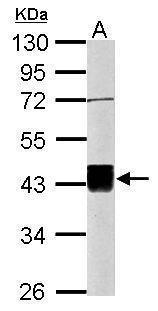
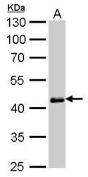
- Main image

- Experimental details
- Sample (20 ?g of whole cell lysate) A: mouse ESC 10% SDS PAGE GTX627419 diluted at 1:1000 The HRP-conjugated anti-mouse IgG antibody (GTX213111-01) was used to detect the primary antibody.
- Submitted by
- GeneTex (provider)
- Main image
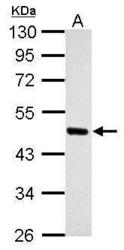
- Experimental details
- Sample (30 ?g of whole cell lysate) A: NT2D1 10% SDS PAGE GTX627419 diluted at 1:1000 The HRP-conjugated anti-mouse IgG antibody (GTX213111-01) was used to detect the primary antibody.

- Submitted by
- GeneTex (provider)
- Main image
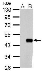
- Experimental details
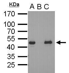
- Sample (10 ?g of whole cell lysate) A: Non-transfected HeLa lysates B: HeLa transfected recombinant Oct4 protein 10% SDS PAGE GTX627419 diluted at 1:1000 The HRP-conjugated anti-mouse IgG antibody (GTX213111-01) was used to detect the primary antibody.
- Submitted by
- GeneTex (provider)
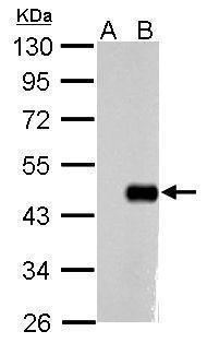
- Main image
- Experimental details
- Oct4 antibody [GT486] detects Oct4 protein by western blot analysis.A. 30 ?g human ESC whole cell lysate/extract10% SDS-PAGE (GTX627419) dilution: 1:1000 The HRP-conjugated anti-mouse IgG antibody (GTX213111-01) was used to detect the primary antibody.

Supportive validation
- Submitted by
- GeneTex (provider)
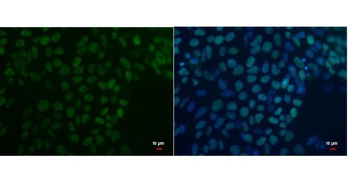
- Main image
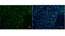
- Experimental details
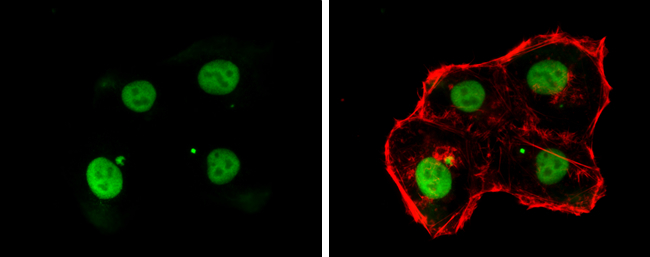
- Oct4 antibody detects Oct4 protein by immunofluorescent analysis. Sample: human embryonic stem cell were fixed in 4% paraformaldehyde for 15 min. Green: Oct4 protein stained by Oct4 antibody (GTX627419) diluted at 1:200. Blue: Hoechst 33342 staining. Scale bar = 10 £gm.
- Submitted by
- GeneTex (provider)
- Main image
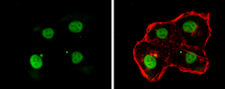
- Experimental details
- Oct4 antibody [GT486] detects Oct4 protein at nucleus by immunofluorescent analysis.Sample: NT2D1 cells were fixed in 4% paraformaldehyde at RT for 15 min.Green: Oct4 stained by Oct4 antibody [GT486] (GTX627419) diluted at 1:500.Red: phalloidin, a cytoskeleton? marker, diluted at 1:100.
Supportive validation
- Submitted by
- GeneTex (provider)
- Main image

- Experimental details
- Oct4 antibody immunoprecipitates Oct4 protein in IP experiments. IP Sample: cell lysate/extract of Oct4 gene transfected 293T cells A. Cell lysate/extract of transfected 293T cell B. Control with 2 £gg of preimmune mouse IgG C. Immunoprecipitation of Oct4 by 2 £gg of Oct4 antibody (GTX627419) 12% SDS-PAGE The immunoprecipitated Oct4 protein was detected by Oct4 antibody (GTX627419) diluted at 1:1000. EasyBlot anti-mouse IgG (GTX221667-01) was used as a secondary reagent.
Supportive validation
- Submitted by
- GeneTex (provider)
- Main image
- Experimental details
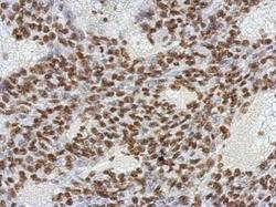
- Immunohistochemical analysis of paraffin-embedded BT483 xenograft, using OCT3/4(GTX627419) antibody at 1:200 dilution.

Supportive validation
- Submitted by
- GeneTex (provider)
- Main image
- Experimental details
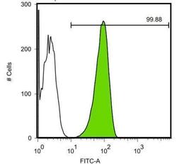
- Oct4 antibody [GT486] detects POU5F protein by flow cytomertry analysis. Sample: Human embryonic stem cells Black: Isotype control dilution: 1:50 Green: Oct4 antibody [GT486] dilution: 1:50
