Explore
 Validate
 Learn
Learn Western blot
Antibody data
- Antibody Data
- Antigen structure
- References [1]
- Comments [0]
- Validations
- Western blot [1]
- Immunocytochemistry [1]
- Flow cytometry [1]
Submit
Validation data
Reference
Comment
Report error
- Product number
- MAB17591 - Provider product page

- Provider
- R&D Systems
- Product name
- Human Oct-4A Antibody
- Antibody type
- Monoclonal
- Description
- Protein A or G purified from hybridoma culture supernatant. Detects human Oct-4A in Western blots.
- Reactivity
- Human
- Host
- Mouse
- Conjugate
- Unconjugated
- Antigen sequence
NP_002692- Isotype
- IgG
- Antibody clone number
- 653108
- Vial size
- 100 ug
- Concentration
- LYOPH
- Storage
- Use a manual defrost freezer and avoid repeated freeze-thaw cycles. 12 months from date of receipt, -20 to -70 °C as supplied. 1 month, 2 to 8 °C under sterile conditions after reconstitution. 6 months, -20 to -70 °C under sterile conditions after reconstitution.
Submitted references Genome-Wide Transcriptome and Binding Sites Analyses Identify Early FOX Expressions for Enhancing Cardiomyogenesis Efficiency of hESC Cultures.
Yeo HC, Ting S, Brena RM, Koh G, Chen A, Toh SQ, Lim YM, Oh SK, Lee DY
Scientific reports 2016 Aug 9;6:31068
Scientific reports 2016 Aug 9;6:31068
No comments: Submit comment
Supportive validation
- Submitted by
- R&D Systems (provider)
- Main image
- Experimental details
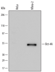
- Detection of Human Oct-4A by Western Blot. Western blot shows lysates of HeLa human cervical epithelial carcinoma cell line and NTera-2 human testicular embryonic carcinoma cell line. PVDF Membrane was probed with 0.5 µg/mL of Mouse Anti-Human Oct-4A Monoclonal Antibody (Catalog # MAB17591) followed by HRP-conjugated Anti-Mouse IgG Secondary Antibody (Catalog # HAF007). A specific band was detected for Oct-4A at approximately 50 kDa (as indicated). This experiment was conducted under reducing conditions and using Immunoblot Buffer Group 1.

Supportive validation
- Submitted by
- R&D Systems (provider)
- Main image

- Experimental details
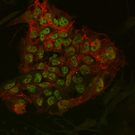
- Oct-4A and E-Cadherin in BG01V Human Stem Cells. Oct-4A and E-Cadherin were detected in human BG01V embryonic stem cells grown on irradiated MEF cells using 10 µg/mL Human Oct-4A Monoclonal Antibody (Catalog # MAB17591) and 10 µg/mL Human E-Cadherin Affinity-purified Polyclonal Antibody (Catalog # AF648). Cells were incubated with primary antibodies for 3 hours at room temperature. Cells were stained for Oct-4A using the Northern-Lights™ 493-conjugated Anti-Mouse IgG Secondary Antibody (green; Catalog # NL009), and stained for E-Cadherin using the Northern-Lights™ 557-conjugated Anti-Goat IgG Secondary Antibody (red; Catalog # NL001). Specific staining of Oct-4A was localized to nuclei. View our protocol for Fluorescent ICC Staining of Cells on Coverslips.
Supportive validation
- Submitted by
- R&D Systems (provider)
- Main image

- Experimental details
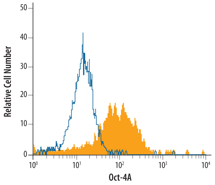
- Detection of Oct-4A in BG01V Human Stem Cells by Flow Cytometry. BG01V human embryonic stem cells was stained with Human Oct-4A Monoclonal Antibody (Catalog # MAB17591, filled histogram) or isotype control antibody (Catalog # MAB003, open histogram), followed by Allophycocyanin-conjugated Anti-Mouse IgG F(ab')2 Secondary Antibody (Catalog # F0101B). To facilitate intracellular staining, cells were fixed with paraformaldehyde and permeabilized with saponin.