 Explore
 Validate
 Learn
Learn Immunoprecipitation
 Flow cytometry
Antibody data
- Antibody Data
- Antigen structure
- References [0]
- Comments [0]
- Validations
- Flow cytometry [1]
- Other assay [4]
Submit
Validation data
Reference
Comment
Report error
- Product number
- 14-5788-80 - Provider product page

- Provider
- Invitrogen Antibodies
- Product name
- MICA/B Monoclonal Antibody (6D4), eBioscience™
- Antibody type
- Monoclonal
- Antigen
- Other
- Description
- Description: The 6D4 monoclonal antibody reacts with the human major histocompatibility complex (MHC) class I chain-related (MIC), MICA and MICB proteins. MICA and MICB are related proteins of 83% amino acid similarity, and show homology with classical human leukocyte antigen (HLA) molecules. The structure of MICA and MICB are similar to classical HLA class I chains, however they do not bind beta2 microglobulin or bind peptide typical of HLA class I. MICA and MICB are expressed on the cell surface of endothelial cells, fibroblasts, gastric epithelium and PHA-stimulated T cells, and act as a ligand for NKGD2 expressed on the surface of NK cells, gammadelta T cells and alpha beta CD8+ T cells. There is evidence to suggest that human cytomegalovirus (HCMV) subverts NK cell detection by inhibiting the function of MICB. Furthermore, MICA and MICB expression has been detected in several epithelial tumours isolated from breast, lung, ovary, prostate, colon and kidney. Applications Reported: This 6D4 antibody has been reported for use in flow cytometric analysis, immunoprecipitation, and immunohistochemical staining. (Please use Functional Grade purified 6D4, Product # 16-5788, in functional assays). Applications Tested: This 6D4 antibody has been tested by flow cytometric analysis of HeLa cells. This can be used at less than or equal to 0.5 µg per test. A test is defined as the amount (µg) of antibody that will stain a cell sample in a final volume of 100 µL. Cell number should be determined empirically but can range from 10^5 to 10^8 cells/test. It is recommended that the antibody be carefully titrated for optimal performance in the assay of interest. Purity: Greater than 90%, as determined by SDS-PAGE. Aggregation: Less than 10%, as determined by HPLC. Filtration: 0.2 µm post-manufacturing filtered.
- Reactivity
- Human
- Host
- Mouse
- Isotype
- IgG
- Antibody clone number
- 6D4
- Vial size
- 25 µg
- Concentration
- 0.5 mg/mL
- Storage
- 4° C
No comments: Submit comment
Supportive validation
- Submitted by
- Invitrogen Antibodies (provider)
- Main image
- Experimental details
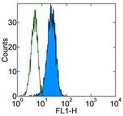
- Staining of HeLa cell line with 0.25 µg of Mouse IgG2a kappa Isotype Control Purified (Product # 14-4724-82) (open histogram) or 0.25 µg of Anti-Human MICA/B Purified (filled histogram) followed by Anti-Mouse IgG FITC (Product # 11-4011-85). Total viable cells were used for analysis.

Supportive validation
- Submitted by
- Invitrogen Antibodies (provider)
- Main image
- Experimental details
- NULL
- Submitted by
- Invitrogen Antibodies (provider)
- Main image
- Experimental details
- NULL
- Submitted by
- Invitrogen Antibodies (provider)
- Main image
- Experimental details
- NULL

- Submitted by
- Invitrogen Antibodies (provider)
- Main image
- Experimental details
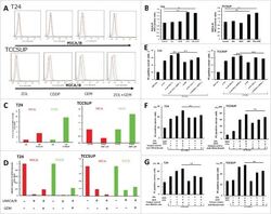
- Figure 4. Changes of MICA/B expression in UBC cells following treatment with gemcitabine and in vitro cytotoxicity assay in MICA/B-knockdown UBC cells. (A) MICA/B expression in T24 and TCCSUP cells before and after ZOL, cisplatin, gemcitabine, and both ZOL and gemcitabine treatment. All agents were used at a concentration of 5 muM for 24 h. MICA/B expression was examined by FACS. A representative histogram is shown. Red histogram: background control; blue: no treatment; orange: agent treatment. (B) Median fluorescence intensity (MFI) of MICA/B expression in each agent-treated sample was quantified and normalized to that of the untreated control sample. Data represent the mean +- SD of triplicate wells. (C) T24 and TCCSUP cells were treated with 5 muM gemcitabine for 24 h. MICA and MICB mRNA transcripts were examined by quantitative RT-PCR. (D) Effects of MICA/B small interfering RNA (siMICA/B). (E) In vitro cytotoxicity assay was performed on MICA/B-knockdown UBC cells. (F) In vitro cytotoxicity assay was performed on UBC cells in the presence of anti-MICA/B blocking mAb or isotype control. (G) In vitro cytotoxicity assay was performed on UBC cells in the presence of anti-NKG2D blocking mAb or isotype control. Data represent the mean +- SD of triplicate wells. Statistical significance is displayed as ** for P < 0.01.
