Explore
 Validate
 Learn
LearnMAB0109
antibody from Abnova Corporation
Targeting: TLR5
FLJ10052, MGC126430, MGC126431, SLEB1, TIL3
 Western blot
Antibody data
- Antibody Data
- Antigen structure
- References [5]
- Comments [0]
- Validations
- Western blot [1]
- Immunohistochemistry [1]
- Flow cytometry [2]
Submit
Validation data
Reference
Comment
Report error
- Product number
- MAB0109 - Provider product page

- Provider
- Abnova Corporation
- Proper citation
- Abnova Corporation Cat#MAB0109, RRID:AB_1017039
- Product name
- TLR5 monoclonal antibody, clone 19D759.2
- Antibody type
- Monoclonal
- Description
- Mouse monoclonal antibody raised against synthetic peptide of TLR5.
- Isotype
- IgG
- Antibody clone number
- 19D759.2
- Storage
- Store at 4°C. For long term storage store at -20°C.Aliquot to avoid repeated freezing and thawing.
Submitted references Stimulation by TLR5 modulates osteoclast differentiation through STAT1/IFN-beta.
Intracellular signaling mechanisms regulating toll-like receptor-mediated activation of eosinophils.
TGF-alpha regulates TLR expression and function on epidermal keratinocytes.
Inhibition of neutrophil apoptosis by TLR agonists in whole blood: involvement of the phosphoinositide 3-kinase/Akt and NF-kappaB signaling pathways, leading to increased levels of Mcl-1, A1, and phosphorylated Bad.
Human CD4+ T cells express TLR5 and its ligand flagellin enhances the suppressive capacity and expression of FOXP3 in CD4+CD25+ T regulatory cells.
Ha H, Lee JH, Kim HN, Kwak HB, Kim HM, Lee SE, Rhee JH, Kim HH, Lee ZH
Journal of immunology (Baltimore, Md. : 1950) 2008 Feb 1;180(3):1382-9
Journal of immunology (Baltimore, Md. : 1950) 2008 Feb 1;180(3):1382-9
Intracellular signaling mechanisms regulating toll-like receptor-mediated activation of eosinophils.
Wong CK, Cheung PF, Ip WK, Lam CW
American journal of respiratory cell and molecular biology 2007 Jul;37(1):85-96
American journal of respiratory cell and molecular biology 2007 Jul;37(1):85-96
TGF-alpha regulates TLR expression and function on epidermal keratinocytes.
Miller LS, Sørensen OE, Liu PT, Jalian HR, Eshtiaghpour D, Behmanesh BE, Chung W, Starner TD, Kim J, Sieling PA, Ganz T, Modlin RL
Journal of immunology (Baltimore, Md. : 1950) 2005 May 15;174(10):6137-43
Journal of immunology (Baltimore, Md. : 1950) 2005 May 15;174(10):6137-43
Inhibition of neutrophil apoptosis by TLR agonists in whole blood: involvement of the phosphoinositide 3-kinase/Akt and NF-kappaB signaling pathways, leading to increased levels of Mcl-1, A1, and phosphorylated Bad.
François S, El Benna J, Dang PM, Pedruzzi E, Gougerot-Pocidalo MA, Elbim C
Journal of immunology (Baltimore, Md. : 1950) 2005 Mar 15;174(6):3633-42
Journal of immunology (Baltimore, Md. : 1950) 2005 Mar 15;174(6):3633-42
Human CD4+ T cells express TLR5 and its ligand flagellin enhances the suppressive capacity and expression of FOXP3 in CD4+CD25+ T regulatory cells.
Crellin NK, Garcia RV, Hadisfar O, Allan SE, Steiner TS, Levings MK
Journal of immunology (Baltimore, Md. : 1950) 2005 Dec 15;175(12):8051-9
Journal of immunology (Baltimore, Md. : 1950) 2005 Dec 15;175(12):8051-9
No comments: Submit comment
Supportive validation
- Submitted by
- Abnova Corporation (provider)
- Main image

- Experimental details
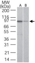
- Western blot analysis of TLR5 in A) Ramos and B) Raw cell lysate using TLR5 monoclonal antibody, clone 19D759.2 (Cat # MAB0109) at 2 ug/mL .
Supportive validation
- Submitted by
- Abnova Corporation (provider)
- Main image
- Experimental details
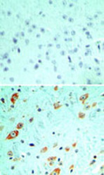
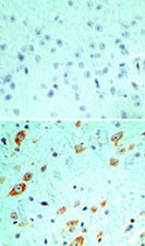
- Immunohistochemical analysis of TLR5 in formalin-fixed, paraffin-embedded mouse brain tissue using an isotype control (top) and TLR5 monoclonal antibody, clone 19D759.2 (Cat # MAB0109) (bottom) at 5 ug/mL .
- Validation comment
- Immunohistochemistry (Formalin/PFA-fixed paraffin-embedded sections)
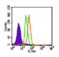
Supportive validation
- Submitted by
- Abnova Corporation (provider)
- Main image

- Experimental details
- Intracellular flow analysis of TLR5 in Ramos cells. Using 1 ug of TLR5 monoclonal antibody, clone 19D759.2 (Cat # MAB0109). Shaded histogram represents cells without antibody; Green represents isotype control (BD Biosciences) ; orange represents TLR5 (Cat # MAB0109) antibody. Secondary antibody Goat Anti-Mouse IgG PE.
- Validation comment
- Flow Cytometry
- Submitted by
- Abnova Corporation (provider)
- Main image

- Experimental details
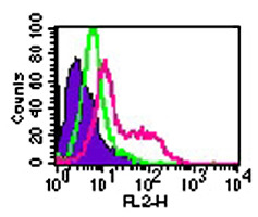
- Intracellular flow analysis of TLR5 in human PBMC (monocytes) using 0.5 ug of TLR5 monoclonal antibody, clone 19D759.2 (Cat # MAB0109). Shaded histogram represents cells without antibody; Green represents isotype control (BD Biosciences) ; red represents TLR5 (Cat # MAB0109). Secondary antibody Goat Anti-Mouse IgG PE.
- Validation comment
- Flow Cytometry