 Explore
 Validate
 Learn
Learn Western blot
 ELISA
ELISAAntibody data
- Antibody Data
- Antigen structure
- References [0]
- Comments [0]
- Validations
- Western blot [1]
- Other assay [3]
Submit
Validation data
Reference
Comment
Report error
- Product number
- 33-9300 - Provider product page

- Provider
- Invitrogen Antibodies
- Product name
- SR Monoclonal Antibody (16H3 (16H3E8))
- Antibody type
- Monoclonal
- Antigen
- Synthetic peptide
- Description
- This monoclonal antibody can be used to detect a sub-set of the non-snRNP splicing factors termed SR proteins. The antibody detects SRp75, SRp55, SRp40, and SRp20 but not SRp30a or b (ASF/SF2, SC-35) proteins. 16H3 also recognizes approximately 20 distinct nuclear proteins including the U1 70K component of the U1 snRNP and both subunits of U2AF. The epitope detected by the 16H3 monoclonal antibody has been termed the “alternating arginine domain” and is composed almost exclusively of argnine alternating with glutamate and aspartate. 16H3 targets are predominantly nuclear, nonnucleolar, and are localized to active sites of polymerase II transcription.
- Reactivity
- Human, Mouse, Rat, Bovine, Canine, Chicken/Avian, Drosophila, Rabbit, Xenopus
- Host
- Mouse
- Isotype
- IgG
- Antibody clone number
- 16H3 (16H3E8)
- Vial size
- 100 µg
- Concentration
- 0.5 mg/mL
- Storage
- Maintain refrigerated at 2-8°C for up to 1 month. For long term storage store at -20°C
No comments: Submit comment
Supportive validation
- Submitted by
- Invitrogen Antibodies (provider)
- Main image
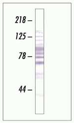
- Experimental details
- Western blot analysis of SR proteins expression in HeLa cell lysate using SR Protein Monoclonal Antibody, Mouse

Supportive validation
- Submitted by
- Invitrogen Antibodies (provider)
- Main image

- Experimental details
- NULL

- Submitted by
- Invitrogen Antibodies (provider)
- Main image

- Experimental details
- NULL

- Submitted by
- Invitrogen Antibodies (provider)
- Main image

- Experimental details
- Figure 1 Increased phosphorylation of SR proteins in PQ-treated cells. A. PQ induced relocalization of SR proteins in the nucleus of treated cells. SH-SY5Y cells treated with vehicle or with 0.75 mM PQ for 18 h were immunostained with an anti-SC35 antibody ( upper row ), transiently transfected with GFP-ASF/SF2 ( middle row ), or with GFP-hnRNPA1 ( lower row ). Nuclei were stained with DAPI. B. Increased phosphorylation of SR proteins in PQ-treated cells. Total extract of control or PQ-treated cells was probed with mAb104 to determine the phosphorylation state of classical SR proteins. C. The same extracts used for the Western blot shown in panel B were probed with the 16H3 monoclonal antibody that detects SR proteins regardless of their phosphorylation status, with anti ASF/SF2 and anti-SRp20 monoclonal antibodies. Actin was used as loading control.
