Explore
 Validate
 Learn
Learn Western blot
Antibody data
- Antibody Data
- Antigen structure
- References [1]
- Comments [0]
- Validations
- Western blot [1]
- Immunohistochemistry [1]
- Flow cytometry [1]
Submit
Validation data
Reference
Comment
Report error
- Product number
- AF8277 - Provider product page

- Provider
- Novus Biologicals
- Product name
- Goat Polyclonal Pref-1/DLK1/FA1 Antibody
- Antibody type
- Polyclonal
- Description
- Antigen Affinity-purified. Detects mouse Pref-1/DLK1/FA1 in direct ELISAs and Western blots. In direct ELISAs, approximately 75% cross-reactivity with recombinant human Pref-1 is observed.
- Reactivity
- Mouse
- Host
- Goat
- Conjugate
- Unconjugated
- Isotype
- IgG
- Vial size
- 100 ug
- Concentration
- LYOPH
- Storage
- Use a manual defrost freezer and avoid repeated freeze-thaw cycles. 12 months from date of receipt, -20 to -70 degreesC as supplied. 1 month, 2 to 8 degreesC under sterile conditions after reconstitution. 6 months, -20 to -70 degreesC under sterile conditions after reconstitution.
Submitted references Mesenchymal Precursor Cells in Adult Nerves Contribute to Mammalian Tissue Repair and Regeneration.
Carr MJ, Toma JS, Johnston APW, Steadman PE, Yuzwa SA, Mahmud N, Frankland PW, Kaplan DR, Miller FD
Cell stem cell 2019 Feb 7;24(2):240-256.e9
Cell stem cell 2019 Feb 7;24(2):240-256.e9
No comments: Submit comment
Supportive validation
- Submitted by
- Novus Biologicals (provider)
- Main image

- Experimental details
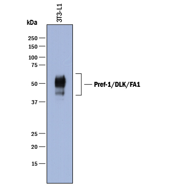
- Detection of Mouse Pref-1/DLK1/FA1 by Western Blot. Western blot shows lysates of 3T3-L1 mouse embryonic fibroblast adipose-like cell line. PVDF membrane was probed with 1 µg/mL of Goat Anti-Mouse Pref-1/DLK1/FA1 Antigen Affinity-purified Polyclonal Antibody (Catalog # AF8277) followed by HRP-conjugated Anti-Goat IgG Secondary Antibody (Catalog # HAF017). A specific band was detected for Pref-1/DLK1/FA1 at approximately 45-60 kDa (as indicated). This experiment was conducted under reducing conditions and using Immunoblot Buffer Group 1.
Supportive validation
- Submitted by
- Novus Biologicals (provider)
- Main image
- Experimental details
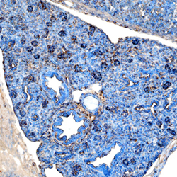
- Pref-1/DLK1/FA1 in Mouse Embryo. Pref-1/DLK1/FA1 was detected in immersion fixed frozen sections of mouse embryonic lung using Goat Anti-Mouse Pref-1/DLK1/FA1 Antigen Affinity-purified Polyclonal Antibody (Catalog # AF8277) at 1 µg/mL overnight at 4 °C. Tissue was stained using the Anti-Goat HRP-DAB Cell & Tissue Staining Kit (brown; Catalog # CTS008) and counterstained with hematoxylin (blue). Specific staining was localized to cytoplasm. View our protocol for Chromogenic IHC Staining of Frozen Tissue Sections.

Supportive validation
- Submitted by
- Novus Biologicals (provider)
- Main image
- Experimental details
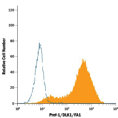
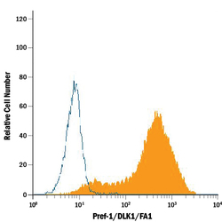
- Detection of Pref-1/DLK1/FA1 in 3T3-L1 Mouse Cell Line by Flow Cytometry. 3T3-L1 mouse embryonic fibroblast adipose-like cell line was stained with Goat Anti-Mouse Pref-1/DLK1/FA1 Antigen Affinity-purified Polyclonal Antibody (Catalog # AF8277, filled histogram) or control antibody (Catalog # AB-108-C, open histogram), followed by Phycoerythrin-conjugated Anti-Goat IgG Secondary Antibody (Catalog # F0107).