 Explore
 Validate
 Learn
Learn Western blot
 Immunohistochemistry
Antibody data
- Antibody Data
- Antigen structure
- References [2]
- Comments [0]
- Validations
- Immunohistochemistry [1]
- Flow cytometry [3]
Submit
Validation data
Reference
Comment
Report error
- Product number
- PA5-72199 - Provider product page

- Provider
- Invitrogen Antibodies
- Product name
- DLK1 Polyclonal Antibody
- Antibody type
- Polyclonal
- Antigen
- Synthetic peptide
- Reactivity
- Human, Mouse
- Host
- Rabbit
- Isotype
- IgG
- Vial size
- 400 μL
- Concentration
- 0.35 mg/mL
- Storage
- Store at 4°C short term. For long term storage, store at -20°C, avoiding freeze/thaw cycles.
Submitted references Hypoxia-induced release, nuclear translocation, and signaling activity of a DLK1 intracellular fragment in glioma.
Niche-derived soluble DLK1 promotes glioma growth.
Grassi ES, Pantazopoulou V, Pietras A
Oncogene 2020 May;39(20):4028-4044
Oncogene 2020 May;39(20):4028-4044
Niche-derived soluble DLK1 promotes glioma growth.
Grassi ES, Jeannot P, Pantazopoulou V, Berg TJ, Pietras A
Neoplasia (New York, N.Y.) 2020 Dec;22(12):689-701
Neoplasia (New York, N.Y.) 2020 Dec;22(12):689-701
No comments: Submit comment
Supportive validation
- Submitted by
- Invitrogen Antibodies (provider)
- Main image
- Experimental details
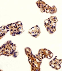
- Immunohistochemistry analysis of DLK1 in paraffin-embedded human placenta section. Samples were incubated with DLK1 polyclonal antibody (Product # PA5-72199) using a dilution of 1:25 followed by an undiluted biotinylated goat polyvalent antibody was used as the secondary, followed by DAB staining.

Supportive validation
- Submitted by
- Invitrogen Antibodies (provider)
- Main image
- Experimental details
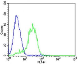
- Flow cytometric analysis of HepG2 cells using DLK1 Antibody (C-term)(green, Product # PA5-72199) compared to an isotype control of mouse IgG2b(blue). AP20959c was diluted at 1:25 dilution. An Alexa Fluor® 488 goat anti-mouse lgG at 1:400 dilution was used as the secondary antibody.

- Submitted by
- Invitrogen Antibodies (provider)
- Main image

- Experimental details
- Flow cytometry analysis of DLK1 in HepG2 cells (green) compared to an isotype control of rabbit IgG (blue). Samples were incubated with DLK1 polyclonal antibody (Product # PA5-72199) using a dilution of 1:25 followed by a Alexa Fluor 488 goat anti-rabbit IgG secondary antibody with a dilution of 1:400.

- Submitted by
- Invitrogen Antibodies (provider)
- Main image

- Experimental details
- Flow cytometry of of DLK1 in HepG2 cells (green). Samples were incubated with DLK1 polyclonal antibody (Product # PA5-72199) using a dilution of 1:25 followed by Alexa Fluor® 488 goat anti-mouse lgG at 1:400. Isotype control of mouse IgG2b (blue).
