Explore
 Validate
 Learn
Learn Western blot
 Immunocytochemistry
 Immunohistochemistry
Antibody data
- Antibody Data
- Antigen structure
- References [3]
- Comments [0]
- Validations
- Western blot [1]
- Immunocytochemistry [1]
Submit
Validation data
Reference
Comment
Report error
- Product number
- HPA011905 - Provider product page

- Provider
- Atlas Antibodies
- Proper citation
- Atlas Antibodies Cat#HPA011905, RRID:AB_1855111
- Product name
- Anti-PDCD6IP
- Antibody type
- Polyclonal
- Description
- Polyclonal Antibody against Human PDCD6IP, Gene description: programmed cell death 6 interacting protein, Alternative Gene Names: AIP1, Alix, Hp95, Validated applications: WB, IHC, ICC, Uniprot ID: Q8WUM4, Storage: Store at +4°C for short term storage. Long time storage is recommended at -20°C.
- Reactivity
- Human
- Host
- Rabbit
- Conjugate
- Unconjugated
- Isotype
- IgG
- Vial size
- 100 µl
- Concentration
- 0.3 mg/ml
- Storage
- Store at +4°C for short term storage. Long time storage is recommended at -20°C.
- Handling
- The antibody solution should be gently mixed before use.
Submitted references En bloc release of MVB‐like small extracellular vesicle clusters by colorectal carcinoma cells
Modulation of paracrine signaling by CD9 positive small extracellular vesicles mediates cellular growth of androgen deprived prostate cancer
Exosomes in colorectal carcinoma formation: ALIX under the magnifying glass
Valcz G, Buzás E, Kittel Á, Krenács T, Visnovitz T, Spisák S, Török G, Homolya L, Zsigrai S, Kiszler G, Antalffy G, Pálóczi K, Szállási Z, Szabó V, Sebestyén A, Solymosi N, Kalmár A, Dede K, Lőrincz P, Tulassay Z, Igaz P, Molnár B
Journal of Extracellular Vesicles 2019;8(1)
Journal of Extracellular Vesicles 2019;8(1)
Modulation of paracrine signaling by CD9 positive small extracellular vesicles mediates cellular growth of androgen deprived prostate cancer
Soekmadji C, Riches J, Russell P, Ruelcke J, McPherson S, Wang C, Hovens C, Corcoran N, Hill M, Nelson C
Oncotarget 2016;8(32):52237-52255
Oncotarget 2016;8(32):52237-52255
Exosomes in colorectal carcinoma formation: ALIX under the magnifying glass
Valcz G, Galamb O, Krenács T, Spisák S, Kalmár A, Patai Á, Wichmann B, Dede K, Tulassay Z, Molnár B
Modern Pathology 2016;29(8):928-938
Modern Pathology 2016;29(8):928-938
No comments: Submit comment
Enhanced validation
- Submitted by
- Atlas Antibodies (provider)
- Enhanced method
- Genetic validation
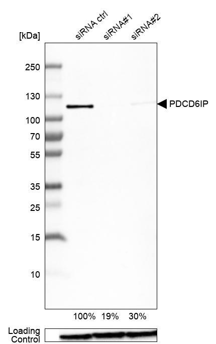
- Main image

- Experimental details
- Western blot analysis in U-138MG cells transfected with control siRNA, target specific siRNA probe #1 and #2, using Anti-PDCD6IP antibody. Remaining relative intensity is presented. Loading control: Anti-GAPDH.
- Sample type
- Human
- Protocol
- Protocol
Supportive validation
- Submitted by
- Atlas Antibodies (provider)
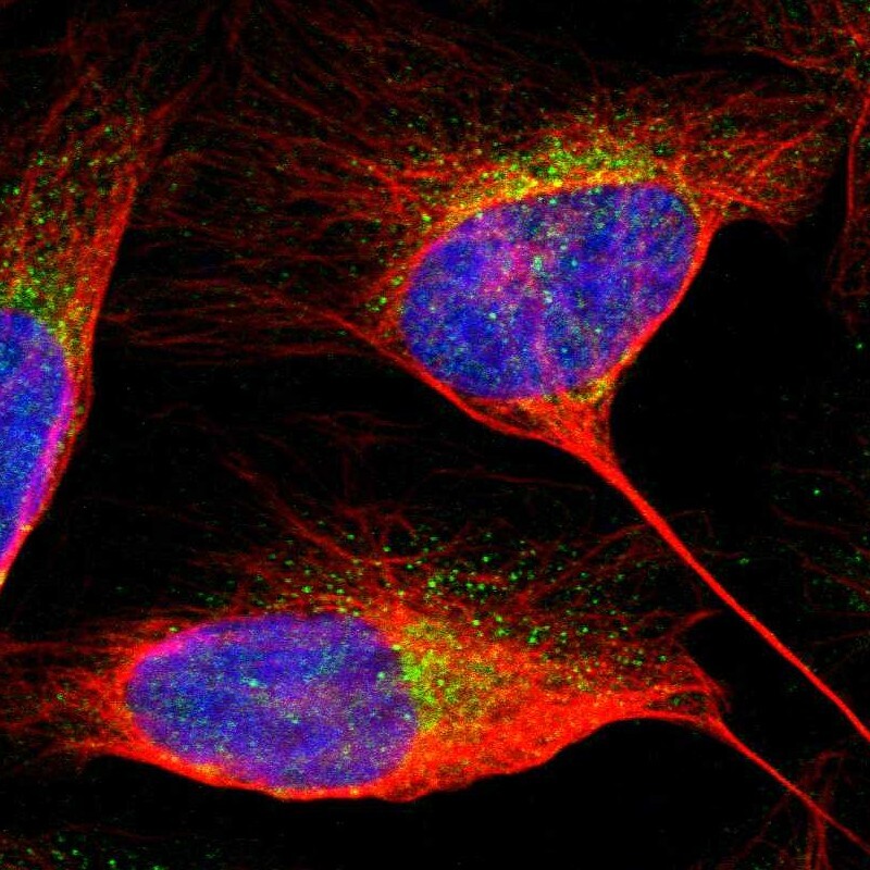
- Main image

- Experimental details
- Immunofluorescent staining of human cell line U-2 OS shows localization to cytosol.
- Sample type
- Human