Explore
 Validate
 Learn
Learn Western blot
 ELISA
ELISA Immunohistochemistry
Antibody data
- Antibody Data
- Antigen structure
- References [0]
- Comments [0]
- Validations
- Western blot [2]
- Immunohistochemistry [6]
Submit
Validation data
Reference
Comment
Report error
- Product number
- LS-C744601 - Provider product page

- Provider
- LSBio
- Product name
- AHSP / EDRF Antibody (Liquid) LS-C744601
- Antibody type
- Polyclonal
- Description
- Delipidated and defibrinated
- Reactivity
- Human, Mouse
- Host
- Rabbit
- Storage
- Store vial at -20°C or below prior to opening. Dilute 1:10 to minimize loss. Store the vial at -20°C or below after dilution. Avoid freeze-thaw cycles.
No comments: Submit comment
Enhanced validation
- Submitted by
- LSBio (provider)
- Enhanced method
- Genetic validation
- Main image
- Experimental details
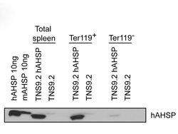
- Western Blot of rabbit anti-AHSP antibody. Lane 1: Recombinant hAHSP (10ng). Lane 2: Recombinant mAHSP (10ng). Lane 3: mice Spleen cells transfected with TNS9.2-hAHSP. Lane 4: mice Spleen cells transfected with TNS9.2 control vector. Lane 5: mice Spleen cells transfected with TNS9.2-hAHSP fractionated by MACS using Ter119+ microbeads. Lane 6: mice Spleen cells transfected with TNS9.2 control vector fractionated by Ter119+. Lane 7: mice Spleen cells transfected with TNS9.2-hAHSP fractionated by Ter119-. Lane 8: Spleen cells from mice transduced with TNS9.2 control vector fractionated by Ter119-. Load: 10 ng per lane. Primary antibody: AHSP antibody at 1:1,000 for overnight at 4°C. Secondary antibody: HRP Streptavidin secondary antibody at 1:40,000 for 30 min at RT. Block: 5% dry milk 30 min at RT. Predicted/Observed size: ~12kDa. Other band(s): none.

- Submitted by
- LSBio (provider)
- Enhanced method
- Genetic validation
- Main image

- Experimental details
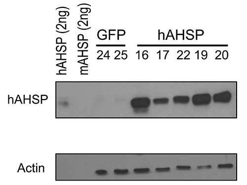
- Western Blot of rabbit anti-AHSP antibody. Lane 1: Recombinant hAHSP (2 ng). Lane 2: Recombinant mAHSP (2 ng). Lane 3: RBC Lysates Mouse #24 - GFP. Lane 4: RBC Lysates Mouse #25 - GFP. Lane 5: RBC Lysates Mouse #16 - hAHSP. Lane 6: RBC Lysates Mouse #17 - hAHSP. Lane 7: RBC Lysates Mouse #22 - hAHSP. Lane 8: RBC Lysates Mouse #19 - hAHSP. Lane 9: RBC Lysates Mouse #20 - hAHSP. Load: if not described differently, 10 ng per lane. Primary antibody: hAHSP antibody, Beta-Actin antibody at 1:1,000 for overnight at 4°C. Secondary antibody: HRP Streptavidin secondary antibody at 1:40,000 for 30 min at RT. Block: 5% dry milk 30 min at RT. Predicted/Observed size: ~12kDa. Other band(s): none.
Supportive validation
- Submitted by
- LSBio (provider)
- Main image
- Experimental details
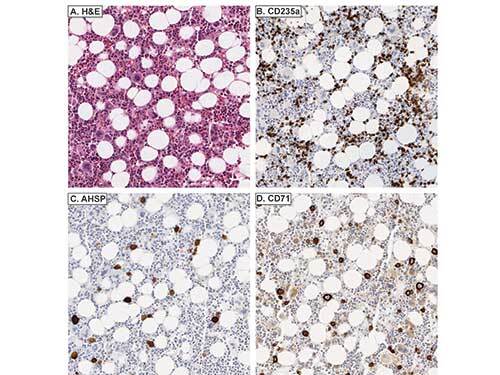
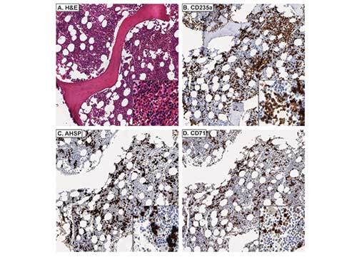
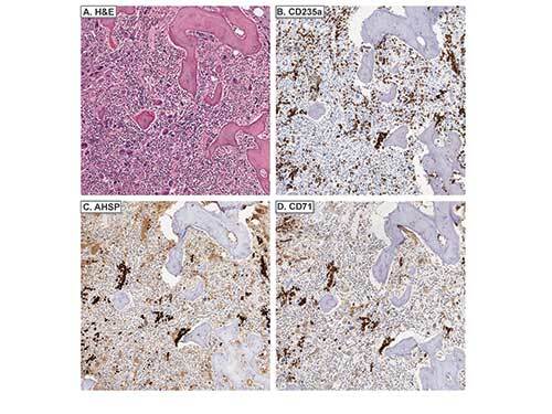
- Immunohistochemistry of rabbit anti-AHSP antibody. Tissue: A. Normal bone marrow, H&E. B. CD235a stains both nucleated EPs and mature, anucleate RBCs. C. AHSP stains nucleated EPs, but not mature, anucleate RBCs. D. CD71 stains nucleated EPs, but not mature, anucleate RBCs. Fixation: acetic acid-zinc-formalin and formalin fixation, embedded in paraffin Antigen retrieval: TRIS-EDTA pH9.0 Primary antibody: AHSP antibody at 1:8,000 for overnight at 4°C Secondary antibody: anti-rabbit secondary at (1:10,000 for 45 min at RT) Localization: Anti-AHSP is cytoplasmic Staining: AHSP antibody as precipitated brown signal with a purple nuclear counterstain using Bond-max – fully automated for IHC.
- Submitted by
- LSBio (provider)
- Main image
- Experimental details
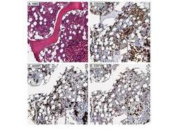
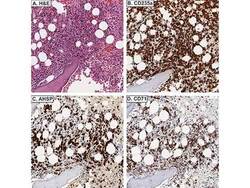
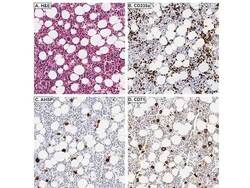
- Immunohistochemistry of rabbit anti-AHSP antibody. Tissue: A. Acute erythroleukemia, H&E. B. CD235a stains erythroid blasts and mature, anucleate RBCs. C. AHSP stains erythroid blasts. D. CD71 stains erythroid blasts. Fixation: acetic acid-zinc-formalin and formalin fixation, embedded in paraffin Antigen retrieval: TRIS-EDTA pH9.0 Primary antibody: AHSP antibody at 1:8,000 for overnight at 4°C Secondary antibody: anti-rabbit secondary at (1:10,000 for 45 min at RT) Localization: Anti-AHSP is cytoplasmic Staining: AHSP antibody as precipitated brown signal with a purple nuclear counterstain using Bond-max – fully automated for IHC.

- Submitted by
- LSBio (provider)
- Main image
- Experimental details
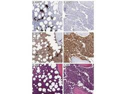
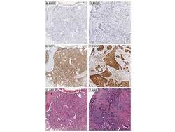
- Immunohistochemistry of rabbit anti-AHSP antibody. Tissue: AHSP stains residual EPs and not myeloid blasts in acute myeloid leukemia with minimal differentiation (A), whereas CD71 stains both myeloid blasts and EPs (B). AHSP does not stain myeloid blasts in acute myelomonocytic leukemia (D), whereas CD71 does (E). C and F are corresponding H&Es, respectively. Fixation: acetic acid-zinc-formalin and formalin fixation, embedded in paraffin Antigen retrieval: TRIS-EDTA pH9.0 Primary antibody: AHSP antibody at 1:8,000 for overnight at 4°C Secondary antibody: anti-rabbit secondary at (1:10,000 for 45 min at RT) Localization: Anti-AHSP is cytoplasmic Staining: AHSP antibody as precipitated brown signal with a purple nuclear counterstain using Bond-max – fully automated for IHC.

- Submitted by
- LSBio (provider)
- Main image
- Experimental details
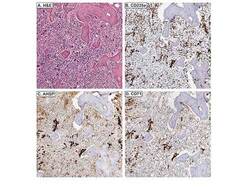
- Immunohistochemistry of rabbit anti-AHSP antibody. Tissue: A. Primary myelofibrosis, H&E. B. CD235a stains both nucleated EPs and mature, anucleate RBCs. AHSP C. and CD71 D. variably stain megakaryocytes and also stain nucleated EPs. Fixation: acetic acid-zinc-formalin and formalin fixation, embedded in paraffin Antigen retrieval: TRIS-EDTA pH9.0 Primary antibody: AHSP antibody at 1:8,000 for overnight at 4°C Secondary antibody: anti-rabbit secondary at (1:10,000 for 45 min at RT) Localization: Anti-AHSP is cytoplasmic Staining: AHSP antibody as precipitated brown signal with a purple nuclear counterstain using Bond-max – fully automated for IHC.
- Submitted by
- LSBio (provider)
- Main image
- Experimental details
- Immunohistochemistry of rabbit anti-AHSP antibody. Tissue: AHSP A. stains residual EPs and not lymphoma cells in DLBCL, whereas CD71 B. stains both lymphomacells and EPs. C. Corresponding H&E. AHSP D. does not metastatic carcinoma, whereas CD71 E. does. F. Corresponding H&E. Fixation: acetic acid-zinc-formalin and formalin fixation, embedded in paraffin Antigen retrieval: TRIS-EDTA pH9.0 Primary antibody: AHSP antibody at 1:8,000 for overnight at 4°C Secondary antibody: anti-rabbit secondary at (1:10,000 for 45 min at RT) Localization: Anti-AHSP is cytoplasmic Staining: AHSP antibody as precipitated brown signal with a purple nuclear counterstain using Bond-max – fully automated for IHC.

- Submitted by
- LSBio (provider)
- Main image
- Experimental details
- Immunohistochemistry of rabbit anti-AHSP antibody. Tissue: Giant pronormoblasts are evident in parvoviral infection (H&E, A). B. CD235a does not stain giant pronormoblasts. AHSP C. and CD71 D. stain giant pronormoblasts. Fixation: acetic acid-zinc-formalin and formalin fixation, embedded in paraffin Antigen retrieval: TRIS-EDTA pH9.0 Primary antibody: AHSP antibody at 1:8,000 for overnight at 4°C Secondary antibody: anti-rabbit secondary at (1:10,000 for 45 min at RT) Localization: Anti-AHSP is cytoplasmic Staining: AHSP antibody as precipitated brown signal with a purple nuclear counterstain using Bond-max – fully automated for IHC.