Explore
 Validate
 Learn
Learn Western blot
 Immunocytochemistry
Antibody data
- Antibody Data
- Antigen structure
- References [1]
- Comments [0]
- Validations
- Immunocytochemistry [2]
- Immunohistochemistry [1]
- Flow cytometry [2]
Submit
Validation data
Reference
Comment
Report error
- Product number
- PA5-25450 - Provider product page

- Provider
- Invitrogen Antibodies
- Product name
- DOPA Decarboxylase Polyclonal Antibody
- Antibody type
- Polyclonal
- Antigen
- Synthetic peptide
- Reactivity
- Human, Mouse
- Host
- Rabbit
- Isotype
- IgG
- Vial size
- 400 μL
- Concentration
- 0.5 mg/mL
- Storage
- Store at 4°C short term. For long term storage, store at -20°C, avoiding freeze/thaw cycles.
Submitted references Gene therapy restores dopamine transporter expression and ameliorates pathology in iPSC and mouse models of infantile parkinsonism.
Ng J, Barral S, De La Fuente Barrigon C, Lignani G, Erdem FA, Wallings R, Privolizzi R, Rossignoli G, Alrashidi H, Heasman S, Meyer E, Ngoh A, Pope S, Karda R, Perocheau D, Baruteau J, Suff N, Antinao Diaz J, Schorge S, Vowles J, Marshall LR, Cowley SA, Sucic S, Freissmuth M, Counsell JR, Wade-Martins R, Heales SJR, Rahim AA, Bencze M, Waddington SN, Kurian MA
Science translational medicine 2021 May 19;13(594)
Science translational medicine 2021 May 19;13(594)
No comments: Submit comment
Supportive validation
- Submitted by
- Invitrogen Antibodies (provider)
- Main image

- Experimental details
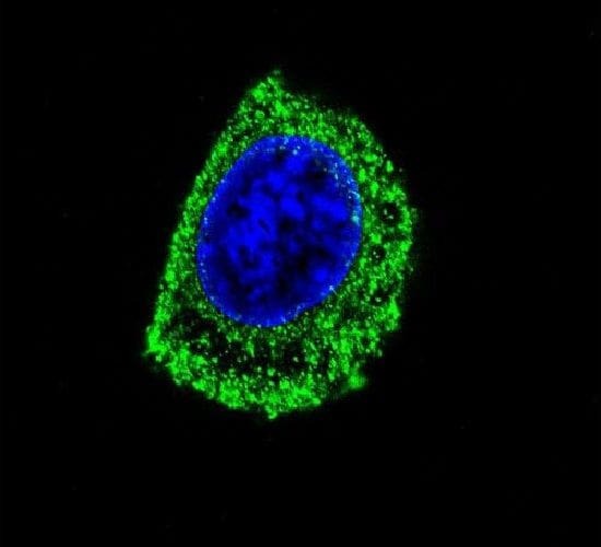
- Immunofluorescent analysis of HepG2 cells using a DDC polyclonal antibody (Product # PA5-25450) at a dilution of 1:10-50, followed by a fluor-conjugated goat anti-rabbit secondary antibody (green). Nuclei were stained with DAPI (blue).
- Submitted by
- Invitrogen Antibodies (provider)
- Main image
- Experimental details
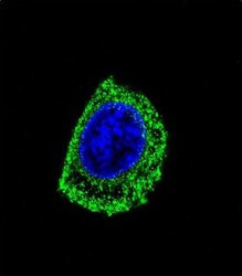
- Immunocytochemistry analysis of DOPA Decarboxylase in HepG2 cells. Samples were incubated in DOPA Decarboxylase polyclonal antibody (Product # PA5-25450) followed by Alexa Fluor® 488-conjugated goat anti-rabbit lgG (green). DAPI was used to stain the cell nuclear (blue).

Supportive validation
- Submitted by
- Invitrogen Antibodies (provider)
- Main image
- Experimental details
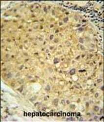
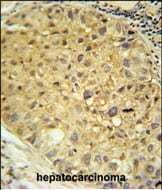
- Immunohistochemistry analysis of DOPA Decarboxylase in formalin fixed and paraffin embedded human hepatocarcinoma. Samples were incubated with DOPA Decarboxylase polyclonal antibody (Product # PA5-25450) followed by peroxidase conjugation of the secondary antibody and DAB staining. This data demonstrates the use of this antibody for immunohistochemistry. Clinical relevance has not been evaluated.
Supportive validation
- Submitted by
- Invitrogen Antibodies (provider)
- Main image
- Experimental details
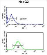
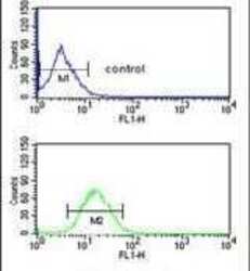
- Flow cytometry analysis of HepG2 cells using a DDC polyclonal antibody (Product # PA5-25450) (bottom) compared to a negative control cell (top) at a dilution of 1:10-50, followed by a FITC-conjugated goat anti-rabbit antibody
- Submitted by
- Invitrogen Antibodies (provider)
- Main image

- Experimental details
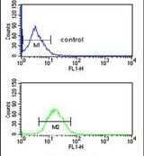
- Flow cytometry of DOPA Decarboxylase in HepG2 cells (bottom histogram). Samples were incubated with DOPA Decarboxylase polyclonal antibody (Product # PA5-25450) followed by FITC-conjugated goat-anti-rabbit secondary antibody. Negative control cell (top histogram).