 Explore
 Validate
 Learn
Learn Western blot
Antibody data
- Antibody Data
- Antigen structure
- References [0]
- Comments [0]
- Validations
- Western blot [1]
- Immunocytochemistry [3]
Submit
Validation data
Reference
Comment
Report error
- Product number
- ABIN2508019 - Provider product page

- Provider
- antibodies-online
- Product name
- anti-Collagen, Type XVIII, alpha 1 (COL18A1) antibody
- Antibody type
- Polyclonal
- Antigen
- Other
- Description
- Produced from sera of rabbits pre-immunized with highly pure (>98%) recombinant hEndostatin (human Endostatin). Anti-Human Endostatin specific antibody was purified by affinity chromatography employing immobilized hEndostatin matrix.
- Reactivity
- Human
- Host
- Rabbit
- Vial size
- 100 μg
- Storage
- -20°C
No comments: Submit comment
Supportive validation
- Submitted by
- antibodies-online (provider)
- Main image
- Experimental details
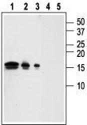
- Western blot analysis of recombinant human GDNF (hGDNF): Lanes 1, 4: 50 ng hGDNF (G-240). Lanes 2, 5: 10 ng hGDNF. Lanes 3, 4 ng hGDNF. Lanes 1-3: Anti-GDNF antibody (ABIN2511166), (1:1000). Lanes 4-5: Anti-GDNF antibody preincubated with the control peptide antigen. Expression of GDNF in rat dorsal root ganglion (DRG). Immunohistochemical staining of rat DRG longitudinal frozen section using Anti-GDNF antibody (ABIN2511166), (1:50), followed by Alexa 555-conjugated goat anti-rabbit antibody (red staining). Hoechst 33342 (blue) was used as a counterstain. GDNF staining is specific for neurons. Note that Schwann cells as well as satellite glial cells are both negative. Expression of GDNF in rat hippocampus Immunohistochemical staining of rat hippocampus frozen sections with Anti-GDNF antibody (ABIN2511166), (1:100), (red). A. GDNF staining is apparent in the pyramidal layer (asterik), in astrocytic fibers (horizontal arrows) and in some interneurons (vertical arrow). B. Calbindin D28k (green) appears in a subset of cells in the pyramidal layer. C. Merge of GDNF and calbindin demonstrates some co-localization in neurons. DAPI is used as the counterstain (blue). Expression of GDNF in rat C6 glioma cells Immunocytochemical staining of GDNF in rat C6 glioma cells. A. Paraformaldehyde-fixed and permeabilized rat C6 glioma cells were stained with Anti-GDNF antibody (ABIN2511166), (1:200) followed by goat anti-rabbit-AlexaFluor-555 secondary antibody (red). Nuclei were visualized with the cell-permeable dye Hoechst 33342 (blue). B. Live view of the same field as in (A).

Supportive validation
- Submitted by
- antibodies-online (provider)
- Main image

- Experimental details
- Image(s): Immunofluorescence

- Submitted by
- antibodies-online (provider)
- Main image

- Experimental details
- Image(s): Immunofluorescence

- Submitted by
- antibodies-online (provider)
- Main image

- Experimental details
- Image(s): Immunofluorescence
