Explore
 Validate
 Learn
LearnPA5-115541
antibody from Invitrogen Antibodies
Targeting: MAP3K8
c-COT, COT, EST, ESTF, MEKK8, Tpl-2
 Western blot
Antibody data
- Antibody Data
- Antigen structure
- References [0]
- Comments [0]
- Validations
- Western blot [1]
- Immunocytochemistry [1]
- Immunohistochemistry [10]
Submit
Validation data
Reference
Comment
Report error
- Product number
- PA5-115541 - Provider product page

- Provider
- Invitrogen Antibodies
- Product name
- TPL2 Polyclonal Antibody
- Antibody type
- Polyclonal
- Antigen
- Synthetic peptide
- Description
- Antibody detects endogenous levels of total MAP3K8/COT.
- Reactivity
- Human, Mouse, Rat
- Host
- Rabbit
- Isotype
- IgG
- Vial size
- 100 μL
- Concentration
- 1 mg/mL
- Storage
- -20°C
No comments: Submit comment
Supportive validation
- Submitted by
- Invitrogen Antibodies (provider)
- Main image
- Experimental details
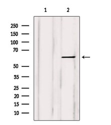
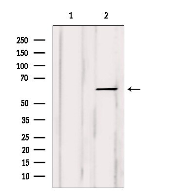
- Western blot analysis of TPL2 in Myeloma cells. Samples were incubated with polyclonal antibody (Product # PA5-115541).
Supportive validation
- Submitted by
- Invitrogen Antibodies (provider)
- Main image
- Experimental details
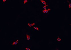
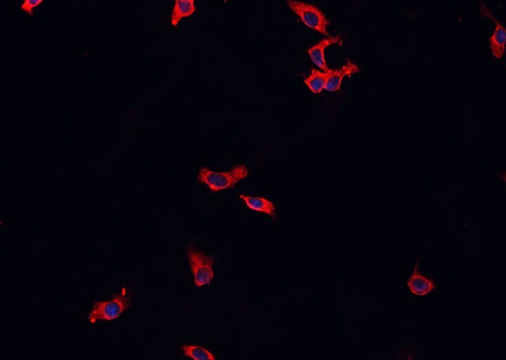
- Immunocytochemistry analysis of TPL2 in HeLa cells. Samples were treated with PFA, permeabilized in 0.1% Triton X-100, blocked in 10% serum (45 min at 25°C), and incubated with polyclonal antibody (Product # PA5-115541) at a dilution of 1:200 (1 hr at 37°C). Secondary staining was applied with Alexa Fluor 594 conjugated goat anti-rabbit IgG (red) using a dilution of 1:600.
Supportive validation
- Submitted by
- Invitrogen Antibodies (provider)
- Main image

- Experimental details
- Immunohistochemistry analysis of TPL2 in rat muscle tissue. Samples were treated with formaldehyde and treated with citrate buffer for antigen retrieval, blocked, and incubated (1.5 hours at 22°C) with polyclonal antibody (Product # PA5-115541) at a dilution of 1:100. Secondary staining was applied with HRP conjugated anti-Rabbit.

- Submitted by
- Invitrogen Antibodies (provider)
- Main image

- Experimental details
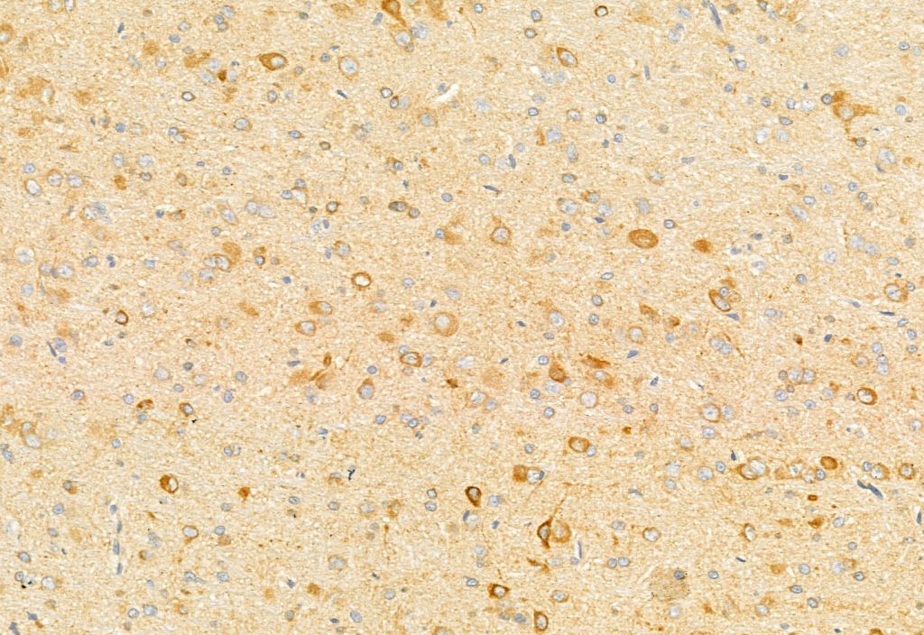
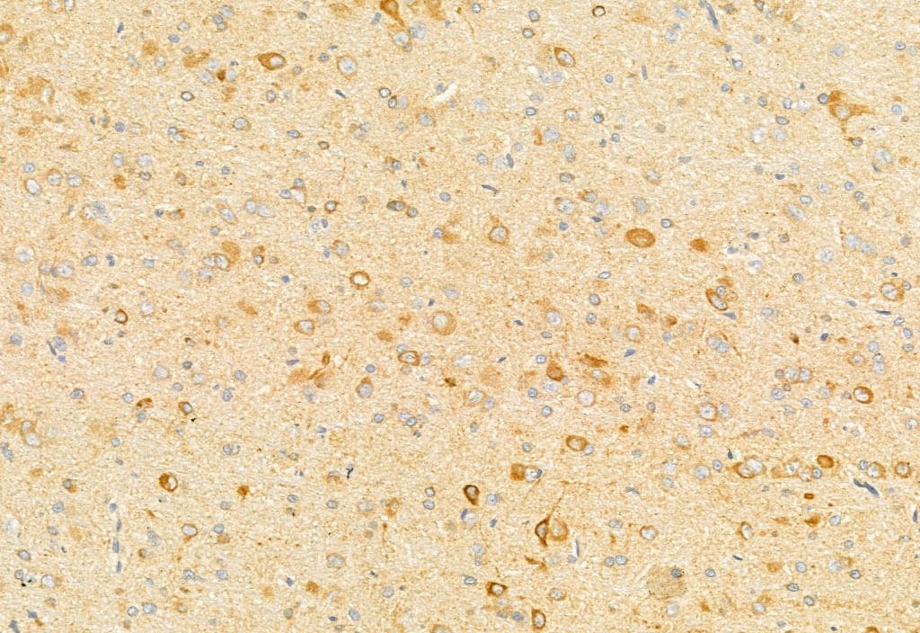
- Immunohistochemistry analysis of TPL2 in rat brain tissue. Samples were treated with formaldehyde and treated with citrate buffer for antigen retrieval, blocked, and incubated (1.5 hours at 22°C) with polyclonal antibody (Product # PA5-115541) at a dilution of 1:100. Secondary staining was applied with HRP conjugated anti-Rabbit.
- Submitted by
- Invitrogen Antibodies (provider)
- Main image

- Experimental details
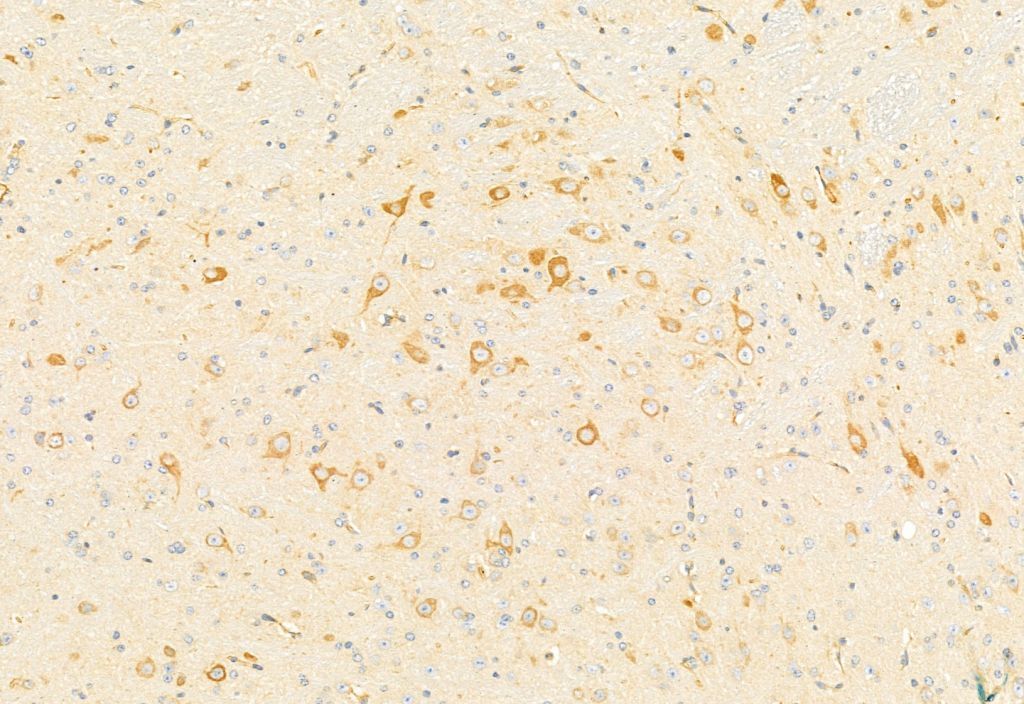
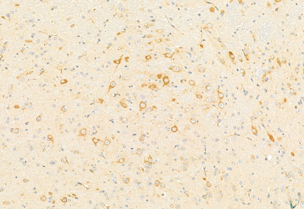
- Immunohistochemistry analysis of TPL2 in mouse brain tissue. Samples were treated with formaldehyde and treated with citrate buffer for antigen retrieval, blocked, and incubated (1.5 hours at 22°C) with polyclonal antibody (Product # PA5-115541) at a dilution of 1:100. Secondary staining was applied with HRP conjugated anti-Rabbit.
- Submitted by
- Invitrogen Antibodies (provider)
- Main image
- Experimental details
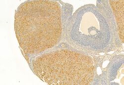
- Immunohistochemistry analysis of TPL2 in rat ovary tissue. Samples were treated with formaldehyde and treated with citrate buffer for antigen retrieval, blocked, and incubated (1.5 hours at 22°C) with polyclonal antibody (Product # PA5-115541) at a dilution of 1:100. Secondary staining was applied with HRP conjugated anti-Rabbit.

- Submitted by
- Invitrogen Antibodies (provider)
- Main image
- Experimental details
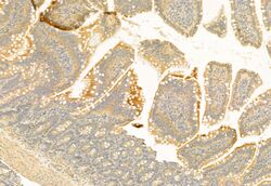
- Immunohistochemistry analysis of TPL2 in rat colon tissue. Samples were treated with formaldehyde and treated with citrate buffer for antigen retrieval, blocked, and incubated (1.5 hours at 22°C) with polyclonal antibody (Product # PA5-115541) at a dilution of 1:100. Secondary staining was applied with HRP conjugated anti-Rabbit.

- Submitted by
- Invitrogen Antibodies (provider)
- Main image
- Experimental details
- Immunohistochemistry analysis of TPL2 in human brain tissue. The sample was formaldehyde fixed and a heat mediated antigen retrieval step in citrate buffer was performed. Samples were incubated with TPL2 polyclonal antibody (Product # PA5-115541) using a dilution of 1:100 (1.5 hours at 22°C) followed by HRP conjugated goat anti-rabbit secondary antibody.

- Submitted by
- Invitrogen Antibodies (provider)
- Main image

- Experimental details
- Immunohistochemistry analysis of TPL2 in rat brain tissue. Samples were treated with formaldehyde and treated with citrate buffer for antigen retrieval, blocked, and incubated (1.5 hours at 22°C) with polyclonal antibody (Product # PA5-115541) at a dilution of 1:100. Secondary staining was applied with HRP conjugated anti-Rabbit.
- Submitted by
- Invitrogen Antibodies (provider)
- Main image

- Experimental details
- Immunohistochemistry analysis of TPL2 in mouse brain tissue. Samples were treated with formaldehyde and treated with citrate buffer for antigen retrieval, blocked, and incubated (1.5 hours at 22°C) with polyclonal antibody (Product # PA5-115541) at a dilution of 1:100. Secondary staining was applied with HRP conjugated anti-Rabbit.
- Submitted by
- Invitrogen Antibodies (provider)
- Main image

- Experimental details
- Immunohistochemistry analysis of TPL2 in rat colon tissue. Samples were treated with formaldehyde and treated with citrate buffer for antigen retrieval, blocked, and incubated (1.5 hours at 22°C) with polyclonal antibody (Product # PA5-115541) at a dilution of 1:100. Secondary staining was applied with HRP conjugated anti-Rabbit.

- Submitted by
- Invitrogen Antibodies (provider)
- Main image
- Experimental details
- Immunohistochemistry analysis of TPL2 in human brain tissue. The sample was formaldehyde fixed and a heat mediated antigen retrieval step in citrate buffer was performed. Samples were incubated with TPL2 polyclonal antibody (Product # PA5-115541) using a dilution of 1:100 (1.5 hours at 22°C) followed by HRP conjugated goat anti-rabbit secondary antibody.