Explore
 Validate
 Learn
Learn Western blot
 Immunoprecipitation
 Immunohistochemistry
Antibody data
- Antibody Data
- Antigen structure
- References [2]
- Comments [0]
- Validations
- Immunohistochemistry [2]
- Other assay [1]
Submit
Validation data
Reference
Comment
Report error
- Product number
- MA1-16739 - Provider product page

- Provider
- Invitrogen Antibodies
- Product name
- Bestrophin 1 Monoclonal Antibody (E6-6)
- Antibody type
- Monoclonal
- Antigen
- Synthetic peptide
- Description
- This antibody does not react with rat samples. Suggested positive control: Pig RPE (retinal Pigment Epithelium) whole cell extract. Immunogen sequence: KDHMDPYWAL ENRDEAHS
- Reactivity
- Human, Mouse, Rat, Canine, Porcine
- Host
- Mouse
- Isotype
- IgG
- Antibody clone number
- E6-6
- Vial size
- 100 μL
- Concentration
- 1.0 mg/mL
- Storage
- -20°C or -80°C if preferred
Submitted references Xeno-free cryopreservation of adherent retinal pigmented epithelium yields viable and functional cells in vitro and in vivo.
A defined subset of clonal retinal stem cell spheres is biased to RPE differentiation.
Pennington BO, Bailey JK, Faynus MA, Hinman C, Hee MN, Ritts R, Nadar V, Zhu D, Mitra D, Martinez-Camarillo JC, Lin TC, Thomas BB, Hinton DR, Humayun MS, Lebkowski J, Johnson LV, Clegg DO
Scientific reports 2021 Mar 18;11(1):6286
Scientific reports 2021 Mar 18;11(1):6286
A defined subset of clonal retinal stem cell spheres is biased to RPE differentiation.
Baakdhah TW, Coles B, van der Kooy D
iScience 2021 Jun 25;24(6):102574
iScience 2021 Jun 25;24(6):102574
No comments: Submit comment
Supportive validation
- Submitted by
- Invitrogen Antibodies (provider)
- Main image
- Experimental details
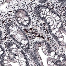
- Immunohistochemical analysis of Bestrophin 1 in immersion fixed paraffin sections of human small intestine. Samples were incubated in Bestrophin 1 monoclonal antibody (Product # MA1-16739) using a dilution of 5 µg/mL for 1 hour at room temperature followed by Anti-Mouse IgG VisUCyte™ HRP Polymer Antibody. Tissue was stained using DAB (brown) and counterstained with hematoxylin (blue). Specific staining was localized to the cell surface and extracellular.

- Submitted by
- Invitrogen Antibodies (provider)
- Main image
- Experimental details
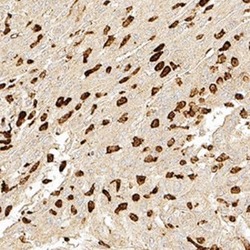
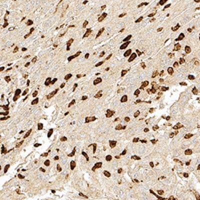
- Immunohistochemical analysis of Bestrophin 1 in immersion fixed paraffin-embedded sections of human brain. Samples were incubated in Bestrophin 1 monoclonal antibody (Product # MA1-16739) using a dilution of 1:300 for 1 hour at room temperature followed by the Anti-Mouse IgG VisUCyte™ HRP Polymer Antibody. Tissue was stained using DAB (brown) and counterstained with hematoxylin (blue). Specific staining was localized to the cytoplasm in neurons.
Supportive validation
- Submitted by
- Invitrogen Antibodies (provider)
- Main image
- Experimental details
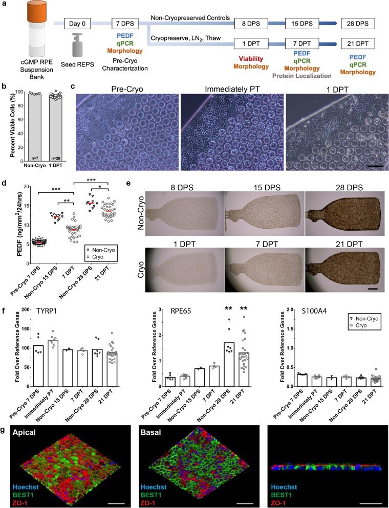
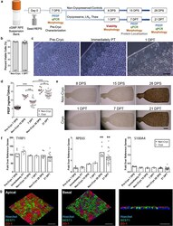
- Figure 2 Cryopreserved REPS retain high viability, cellular identity, and secretory function post-thaw. ( a ) Schematic of overall research design indicates the characterization assays conducted on the designated day post-seeding (DPS) or day post-thaw (DPT). Non-cryopreserved REPS were maintained in culture to provide an age-matched control for each day of sample collection. ( b ) Cryopreserved REPS exhibit 95 +- 2.3% viability as measured by propidium iodide exclusion at 1 DPT (n = 26); viability of non-cryopreserved control REPS (n = 7) averaged to 97 +- 0.89% (mean +- SD., * P < 0.05, unpaired two-tailed t-test). ( c ) REPS exhibit epithelial, cuboidal morphology prior to cryopreservation (Pre-Cryo), immediately post-thaw (PT), and 1 DPT. (Phase contrast, scale bar, 100 um.) ( d ) Amount of secreted PEDF by non-cryopreserved control REPS (black triangles) and cryopreserved/thawed REPS (gray circles) as measured by ELISA. Thawed REPS exhibited a significant increase in secreted PEDF at 7 DPT compared to Pre-Cryopreservation, and also at 21 DPT compared to 7 DPT (*** P < 0.001). A significant difference was detected between thawed REPS and age-matched non-cryopreserved controls at 7 DPT (** P < 0.01) and at 21 DPT (* P < 0.05). (Pre-Cryo, n = 93; Non-Cryo, n = 10; Cryo, n = 33. Each data point represents the mean of two technical replicates. Horizontal red bar indicates mean of the biological replicates. Independent samples Kruskal-Wallis test with pairwise comparisons.) (