Explore
 Validate
 Learn
Learn Flow cytometry
Antibody data
- Antibody Data
- Antigen structure
- References [5]
- Comments [0]
- Validations
- Flow cytometry [1]
- Other assay [1]
Submit
Validation data
Reference
Comment
Report error
- Product number
- 17-6979-42 - Provider product page

- Provider
- Invitrogen Antibodies
- Product name
- CD97 Monoclonal Antibody (VIM3b), APC, eBioscience™
- Antibody type
- Monoclonal
- Antigen
- Other
- Description
- Description: This VIM3b monoclonal antibody reacts with human CD97, a member of the epidermal growth factor (EGF)-TM7 family of cell surface receptors. Present on a variety of hematopoietic cells, CD97 is most highly expressed on granulocytes and monocytes. CD97 can also be detected on lymphocytes, with T and NK cells expressing higher levels than B cells. Moreover, CD97 expression on T lymphocytes can be induced by stimulation of CD3 and CD28. Studies have demonstrated that CD97 is involved in granulocyte trafficking and T cell activation. CD55, also known as Decay-accelerating factor (DAF), is the ligand of CD97. Due to alternative splicing, CD97 exists as three isoforms with molecular weights of 74, 80, and 86 kDa. The VIM3b monoclonal antibody recognizes each form. Applications Reported: This VIM3b antibody has been reported for use in flow cytometric analysis. Applications Tested: This VIM3b antibody has been pre-titrated and tested by flow cytometric analysis of normal human peripheral blood cells. This can be used at 5 µL (1 µg) per test. A test is defined as the amount (µg) of antibody that will stain a cell sample in a final volume of 100 µL. Cell number should be determined empirically but can range from 10^5 to 10^8 cells/test. Excitation: 633-647 nm; Emission: 660 nm; Laser: Red Laser. Filtration: 0.2 µm post-manufacturing filtered.
- Reactivity
- Human
- Host
- Mouse
- Isotype
- IgG
- Antibody clone number
- VIM3b
- Vial size
- 100 Tests
- Concentration
- 5 μL/Test
- Storage
- 4°C, store in dark, DO NOT FREEZE!
Submitted references Quantitative Proteomics Analysis of Lytic KSHV Infection in Human Endothelial Cells Reveals Targets of Viral Immune Modulation.
The cell surface proteome of human mesenchymal stromal cells.
Differential expression of CD97 on human lymphocyte subsets and limited effect of CD97 antibodies on allogeneic T-cell stimulation.
Costimulation via CD55 on human CD4+ T cells mediated by CD97.
CD97 isoform expression in leukocytes.
Gabaev I, Williamson JC, Crozier TWM, Schulz TF, Lehner PJ
Cell reports 2020 Oct 13;33(2):108249
Cell reports 2020 Oct 13;33(2):108249
The cell surface proteome of human mesenchymal stromal cells.
Niehage C, Steenblock C, Pursche T, Bornhäuser M, Corbeil D, Hoflack B
PloS one 2011;6(5):e20399
PloS one 2011;6(5):e20399
Differential expression of CD97 on human lymphocyte subsets and limited effect of CD97 antibodies on allogeneic T-cell stimulation.
Kop EN, Matmati M, Pouwels W, Leclercq G, Tak PP, Hamann J
Immunology letters 2009 Apr 27;123(2):160-8
Immunology letters 2009 Apr 27;123(2):160-8
Costimulation via CD55 on human CD4+ T cells mediated by CD97.
Capasso M, Durrant LG, Stacey M, Gordon S, Ramage J, Spendlove I
Journal of immunology (Baltimore, Md. : 1950) 2006 Jul 15;177(2):1070-7
Journal of immunology (Baltimore, Md. : 1950) 2006 Jul 15;177(2):1070-7
CD97 isoform expression in leukocytes.
Eichler W
Journal of leukocyte biology 2000 Oct;68(4):561-7
Journal of leukocyte biology 2000 Oct;68(4):561-7
No comments: Submit comment
Supportive validation
- Submitted by
- Invitrogen Antibodies (provider)
- Main image
- Experimental details
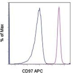
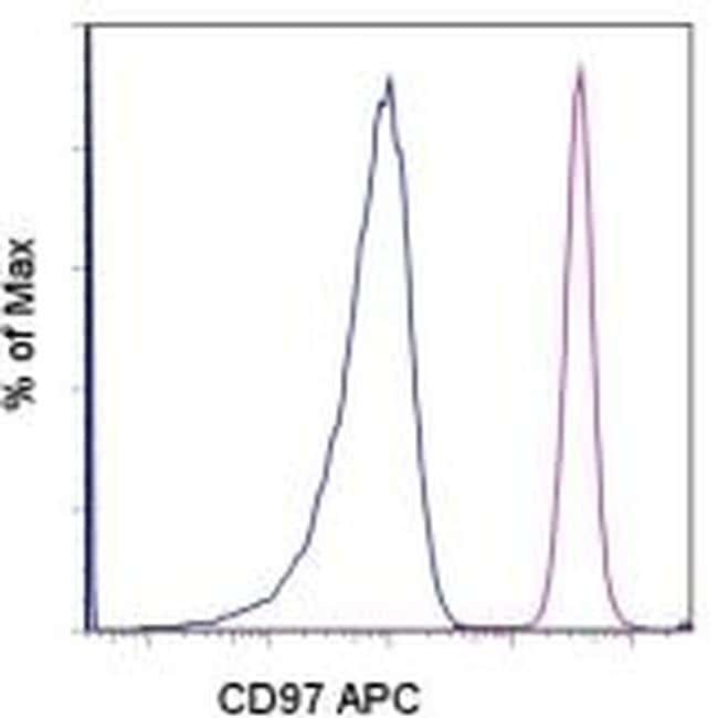
- Staining of normal human peripheral blood cells with Mouse IgG1 kappa Isotype Control APC (Product # 17-4714-81) (blue histogram) or Anti- Human CD97 APC (purple histogram). Cells in the granulocyte gate were used for analysis.
Supportive validation
- Submitted by
- Invitrogen Antibodies (provider)
- Main image
- Experimental details
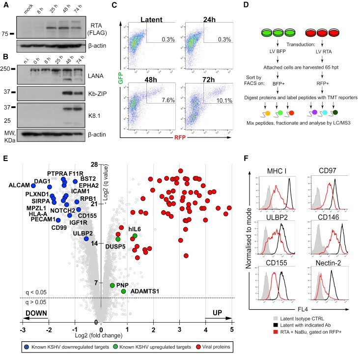
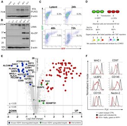
- Figure 1 Lytic KSHV Induces Massive Changes in the Proteome of Endothelial Cells (A and B) Immunoblot analysis of HuAR2T.rKSHV.219 cells transduced with LV RTA, harvested at indicated time points (8, 25, 48, and 74 h), and probed with the indicated antibody. (C) Flow cytometry analysis of HuAR2T.rKSHV.219 cells mock-transduced (latent) or transduced with LV RTA and harvested at the indicated time points (24, 48, and 72 h). (D) Schematic overview of the quantitative proteomics analysis of the cells with latent versus lytic KSHV infection. HuAR2T.rKSHV.219 cells were transduced with LV RTA or control LV BFP, sorted on BFP+ or RFP+, and analyzed by mass spectrometry (MS). (E) Scatterplot displays pairwise comparison between latent and lytic KSHV infection. Each point represents a single protein, plotted by its log2 (fold change in abundance) versus the statistical significance (q value) of that change. Value was corrected for multiple hypothesis testing using the method of Benjamini-Hochberg. Dotted line: q = 0.05. (F) Flow cytometry analysis of HuAR2T.rKSHV.219 Cas9 cells untreated or treated with reactivation mix and stained with the indicated antibody. See also Figure S1 and Table S1 .