Explore
 Validate
 Learn
Learn Western blot
 ELISA
ELISA Immunocytochemistry
Antibody data
- Antibody Data
- Antigen structure
- References [0]
- Comments [0]
- Validations
- Western blot [2]
- Immunocytochemistry [1]
Submit
Validation data
Reference
Comment
Report error
- Product number
- GTX48818 - Provider product page

- Provider
- GeneTex
- Proper citation
- GeneTex Cat#GTX48818, RRID:AB_11175639
- Product name
- CD97 antibody
- Antibody type
- Polyclonal
- Reactivity
- Human, Mouse
- Host
- Rabbit
No comments: Submit comment
Enhanced validation
Supportive validation
- Submitted by
- GeneTex (provider)
- Enhanced method
- Genetic validation
- Main image
- Experimental details
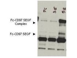
- Western blot using GeneTex's Protein A purified anti-CD97 antibody shows detection of bands corresponding to free Fc-CD97- (5EGF) (lower arrowhead) and Fc-CD97- (5EGF) present as a complex (upper arrowhead) in lysates from COS cells. The left lane contains lysate from cells transfected with control DNA. The right lane contains lysate from COS cells expressing Fc-CD97- (5EGF). Approximately 10 ul of lysate was used in each lane. A 1:1,000 dilution of the primary antibody was used. The image was processed using a 10-sec exposure.
Supportive validation
- Submitted by
- GeneTex (provider)
- Main image

- Experimental details
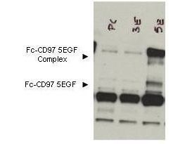
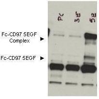
- Western blot using GeneTex's Protein A purified anti-CD97 antibody shows detection of bands corresponding to free Fc-CD97- (5EGF) (lower arrowhead) and Fc-CD97- (5EGF) present as a complex (upper arrowhead) in lysates from COS cells. The left lane contains lysate from cells transfected with control DNA. The right lane contains lysate from COS cells expressing Fc-CD97- (5EGF). No staining was noted from bone marrow lysates taken from CD97 knockout mice. The identity of the band at ~65 kDa appearing in all lanes is not known. The formation of the CD97 complex is currently under investigation. Approximately 10 ul of lysate was used in each lane. A 1:1,000 dilution of the primary antibody was used. The image was processed using a 10-sec exposure. Personal Communication. Yvona Ward. NIH, NCI, CCR, Bethesda, MD.
- Validation comment
- WB
Supportive validation
- Submitted by
- GeneTex (provider)
- Main image

- Experimental details
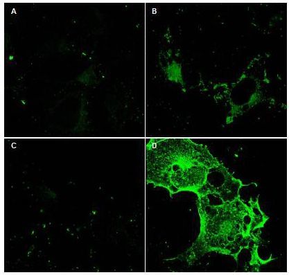
- Immunofluorescence Microscopy using GeneTex Protein A purified anti-CD97 antibody (GTX48818) shows staining of Fc-CD97-(5EGF) (panel D) in transfected COS cells. Panel A and C shows similar staining using pre-immune serum. Panel A and B show staining of mock transfected COS cells (no vector). A 1:2,500 dilution of the primary antibody was used.