Explore
 Validate
 Learn
Learn Immunohistochemistry
Antibody data
- Antibody Data
- Antigen structure
- References [1]
- Comments [0]
- Validations
- Immunohistochemistry [1]
- Other assay [2]
Submit
Validation data
Reference
Comment
Report error
- Product number
- PA5-59485 - Provider product page

- Provider
- Invitrogen Antibodies
- Product name
- HERPUD1 Polyclonal Antibody
- Antibody type
- Polyclonal
- Antigen
- Recombinant full-length protein
- Description
- Immunogen sequence: LGPGFSGYTP YGWLQLSWFQ QIYARQYYMQ YLAATAASGA FVPPPSAQEI PVVSAPAPAP IHNQFPAENQ PANQNAAPQV VVNPGANQNL RMNAQGGPIV EEDDEINRD
- Concentration
- 1 mg/mL
Submitted references Herpud1 suppress angiotensin II induced hypertrophy in cardiomyocytes.
Mikawa M, Sakai C, Yamamoto T, Nakamura Y, Tanaka S, Tominaga N, Inamitsu M, Oda T, Kobayashi S, Yano M
Biochemistry and biophysics reports 2022 Jul;30:101248
Biochemistry and biophysics reports 2022 Jul;30:101248
No comments: Submit comment
Supportive validation
- Submitted by
- Invitrogen Antibodies (provider)
- Main image
- Experimental details
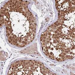
- Immunohistochemical staining of HERPUD1 in human testis tissue shows strong cytoplasmic and nuclear positivity in cells in seminiferus ducts. Samples were probed using a HERPUD1 Polyclonal Antibody (Product # PA5-59485).

Supportive validation
- Submitted by
- Invitrogen Antibodies (provider)
- Main image
- Experimental details
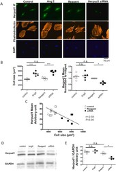
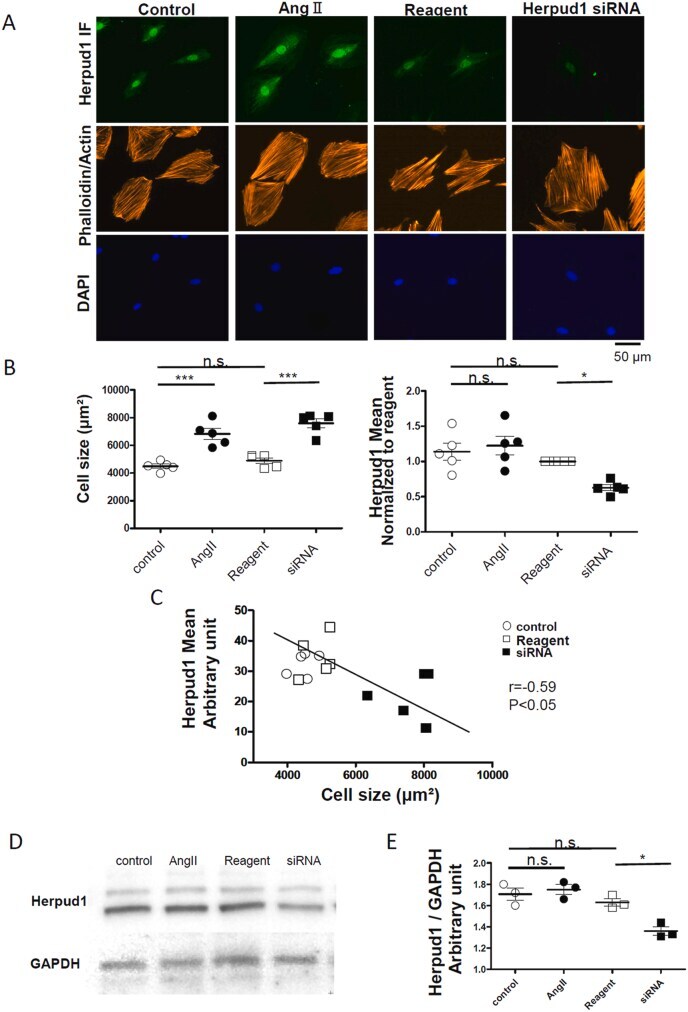
- Silencing Herpud1 resulted in cardiac hypertrophy (A) Representative images of H9C2 cells of homocysteine-responsive endoplasmic reticulum-resident ubiquitin-like domain member 1 (Herpud1) immunofluorescent, phalloidin/actin, and DAPI staining. (B) Summarized data of H9C2 cell size and relative Herpud1 expression normalized to transfection reagent only control. One dot represents mean of 18-58 cells from one experiment. (C) Relation between H9C2 cell size and Herpud1 expression. One dot represents mean of 18-58 cells from one experiment. (D) Representative images of western blotting of whole cell lysate of H9C2 cell by anti-Herpud1 antibody. (E) Summarized data of Herpud 1 protein level in H9C2 cell. *:P < 0.05, ***:P < 0.001. Fig. 1
- Submitted by
- Invitrogen Antibodies (provider)
- Main image

- Experimental details
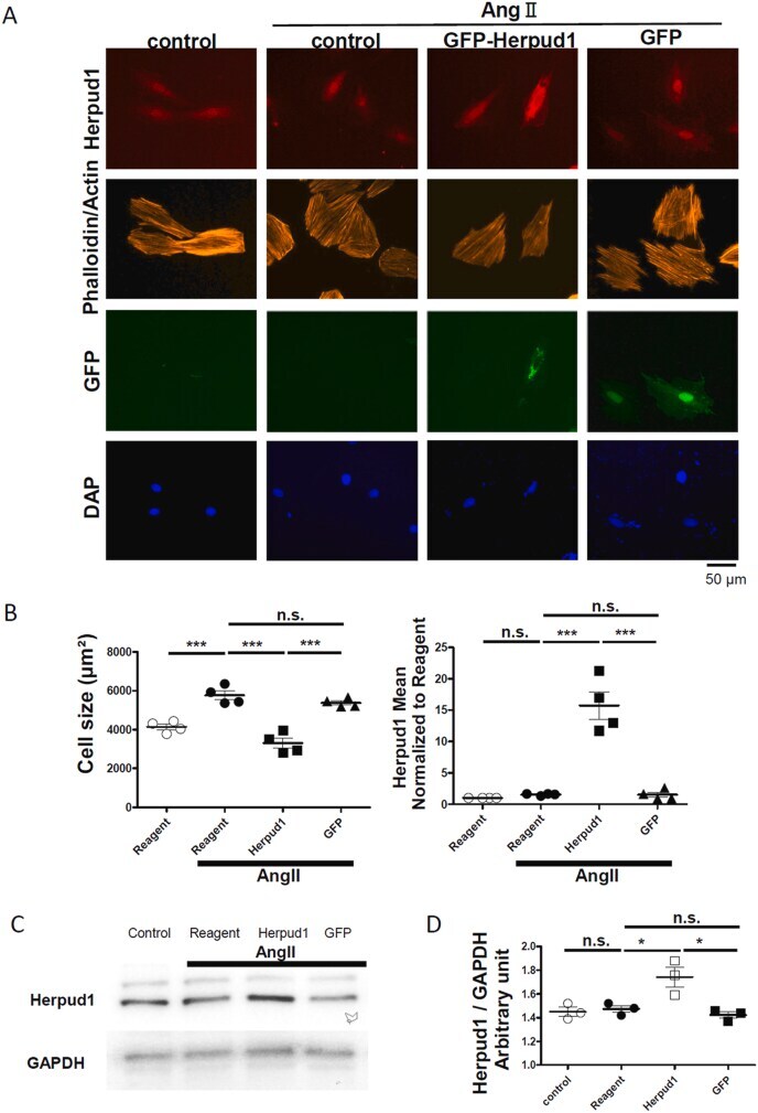
- Overexpression of Herpud1 suppressed the Ang II-induced cardiac hypertrophy (A) Representative images of H9C2 cells of homocysteine-responsive endoplasmic reticulum-resident ubiquitin-like domain member 1 (Herpud1) immunofluorescent, phalloidin/actin, GFP, and DAPI staining. (B) Summarized data of H9C2 cell size and relative Herpud1 expression normalized to transfection reagent only control. One dot represents mean of 7-33 cells from one experiment. (C) Representative images of western blotting of whole cell lysate of H9C2 cell by anti-Herpud1 antibody. (D) Summarized data of Herpud 1 protein level in H9C2 cell. *:P < 0.05, ***:P < 0.001. Fig. 2