Explore
 Validate
 Learn
Learn Western blot
 Immunohistochemistry
Antibody data
- Antibody Data
- Antigen structure
- References [2]
- Comments [0]
- Validations
- Immunohistochemistry [1]
- Other assay [1]
Submit
Validation data
Reference
Comment
Report error
- Product number
- PA5-29469 - Provider product page

- Provider
- Invitrogen Antibodies
- Product name
- HERPUD1 Polyclonal Antibody
- Antibody type
- Polyclonal
- Antigen
- Recombinant full-length protein
- Description
- Recommended positive controls: 293T, mouse brain. Predicted reactivity: Mouse (86%), Rat (83%), Bovine (87%). Store product as a concentrated solution. Centrifuge briefly prior to opening the vial.
- Reactivity
- Human, Mouse
- Host
- Rabbit
- Isotype
- IgG
- Vial size
- 100 μL
- Concentration
- 1 mg/mL
- Storage
- Store at 4°C short term. For long term storage, store at -20°C, avoiding freeze/thaw cycles.
Submitted references Transcriptomic Changes Associated with Loss of Cell Viability Induced by Oxysterol Treatment of a Retinal Photoreceptor-Derived Cell Line: An In Vitro Model of Smith-Lemli-Opitz Syndrome.
Herpud1 negatively regulates pathological cardiac hypertrophy by inducing IP3 receptor degradation.
Pfeffer BA, Xu L, Fliesler SJ
International journal of molecular sciences 2021 Feb 26;22(5)
International journal of molecular sciences 2021 Feb 26;22(5)
Herpud1 negatively regulates pathological cardiac hypertrophy by inducing IP3 receptor degradation.
Torrealba N, Navarro-Marquez M, Garrido V, Pedrozo Z, Romero D, Eura Y, Villalobos E, Roa JC, Chiong M, Kokame K, Lavandero S
Scientific reports 2017 Oct 17;7(1):13402
Scientific reports 2017 Oct 17;7(1):13402
No comments: Submit comment
Supportive validation
- Submitted by
- Invitrogen Antibodies (provider)
- Main image
- Experimental details
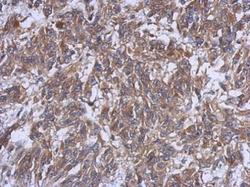
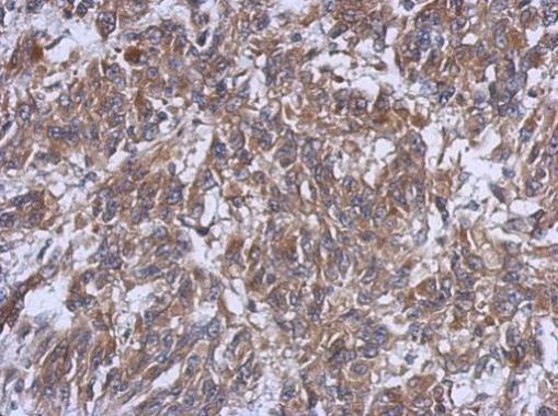
- Immunohistochemical analysis of paraffin-embedded U251 xenograft, using HERPUD1 (Product # PA5-29469) antibody at 1:500 dilution. Antigen Retrieval: EDTA based buffer, pH 8.0, 15 min.
Supportive validation
- Submitted by
- Invitrogen Antibodies (provider)
- Main image

- Experimental details
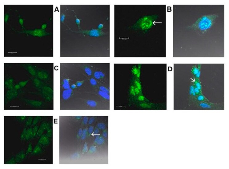
- Figure 19 ( A - E ): Immunolocalization of HERPUD1. ( A - C ), 661W cells were fixed with methacarn; ( D , E ), cells fixed with formaldehyde. ( A , B ): For 8 muM EPCD-treated 661W cells, there were large, dense aggregates of HERPUD1 immunoreactivity (green pseudocolor in left images, and blue-green superimposition with DAPI fluorescence) detected in the nuclear zones (arrow in B ). Bar = 10 mum in ( B ). ( C ): DMSO (VC) substituted for EPCD incubation. Predominantly non-specific background fluorescence, with only sparse, punctate immunoreaction in the vicinity of nuclei. Nuclei exhibit only DAPI staining (blue pseudocolor). ( D ): 661W cells treated with 25 muM 7kCHOL display both nuclear and juxtanuclear (arrow) HERPUD1 immunofluorescent signal, the former as aggregates of various sizes, coinciding partly with DAPI fluorescence, along with less intense, punctate cytoplasmic immunoreactivity. ( E ): Cells incubated with hpbetaCD VC demonstrate a focus of HERPUD1-specific immunofluorescence above background near nuclei (arrow), with some punctate cytoplasmic immunoreactivity; nuclei show primarily DAPI staining only, compared to the 7kCHOL-treated sample ( D ).