Explore
 Validate
 Learn
Learn Western blot
 Immunocytochemistry
Antibody data
- Antibody Data
- Antigen structure
- References [1]
- Comments [0]
- Validations
- Western blot [1]
- Immunocytochemistry [1]
- Immunohistochemistry [1]
Submit
Validation data
Reference
Comment
Report error
- Product number
- AMAb90967 - Provider product page

- Provider
- Atlas Antibodies
- Proper citation
- Atlas Antibodies Cat#AMAb90967, RRID:AB_2665739
- Product name
- Anti-VPS26A
- Antibody type
- Monoclonal
- Description
- Monoclonal Antibody against Human VPS26A, Clone ID: CL2287, Gene description: Vps26, retromer complex component a, Alternative Gene Names: Hbeta58, PEP8A, VPS26, Validated applications: IHC, ICC, WB, Uniprot ID: O75436, Storage: Store at +4°C for short term storage. Long time storage is recommended at -20°C.
- Reactivity
- Human
- Host
- Mouse
- Conjugate
- Unconjugated
- Isotype
- IgG
- Antibody clone number
- CL2287
- Vial size
- 100 µl
- Concentration
- 1.0 mg/ml
- Storage
- Store at +4°C for short term storage. Long time storage is recommended at -20°C.
- Handling
- The antibody solution should be gently mixed before use.
Submitted references BioID reveals an ATG9A interaction with ATG13‐ATG101 in the degradation of p62/SQSTM1‐ubiquitin clusters
Kannangara A, Poole D, McEwan C, Youngs J, Weerasekara V, Thornock A, Lazaro M, Balasooriya E, Oh L, Soderblom E, Lee J, Simmons D, Andersen J
EMBO reports 2021;22(10)
EMBO reports 2021;22(10)
No comments: Submit comment
Enhanced validation
- Submitted by
- Atlas Antibodies (provider)
- Enhanced method
- Genetic validation
- Main image
- Experimental details
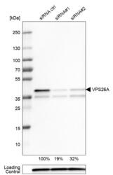
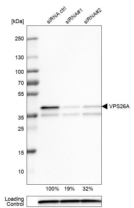
- Western blot analysis in U-251MG cells transfected with control siRNA, target specific siRNA probe #1 and #2, using Anti-VPS26A antibody. Remaining relative intensity is presented. Loading control: Anti-PPIB.
- Sample type
- Human
- Protocol
- Protocol
Supportive validation
- Submitted by
- Atlas Antibodies (provider)
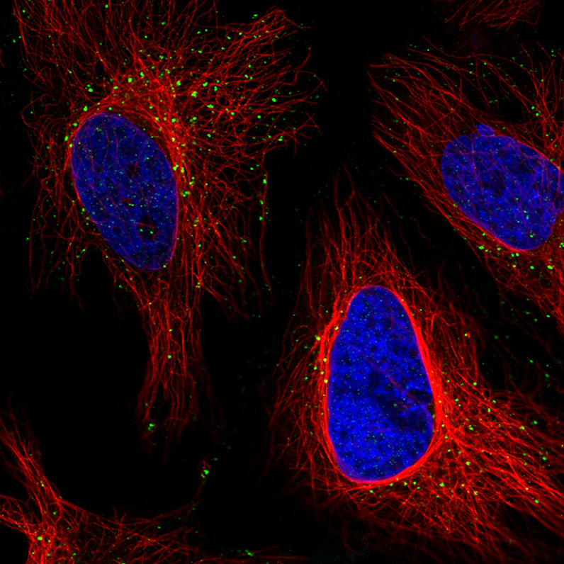
- Main image
- Experimental details
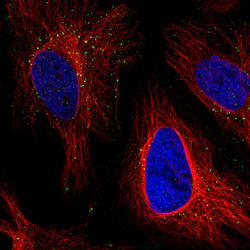
- Immunofluorescence staining in HeLa cell line with Anti-VSP26A monoclonal antibody, showing distinct staining of endosomes in green. Microtubule- and nuclear probes are visualized in red and blue respectively (where available).
- Sample type
- Human
Supportive validation
- Submitted by
- Atlas Antibodies (provider)
- Enhanced method
- Orthogonal validation
- Main image
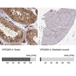
- Experimental details
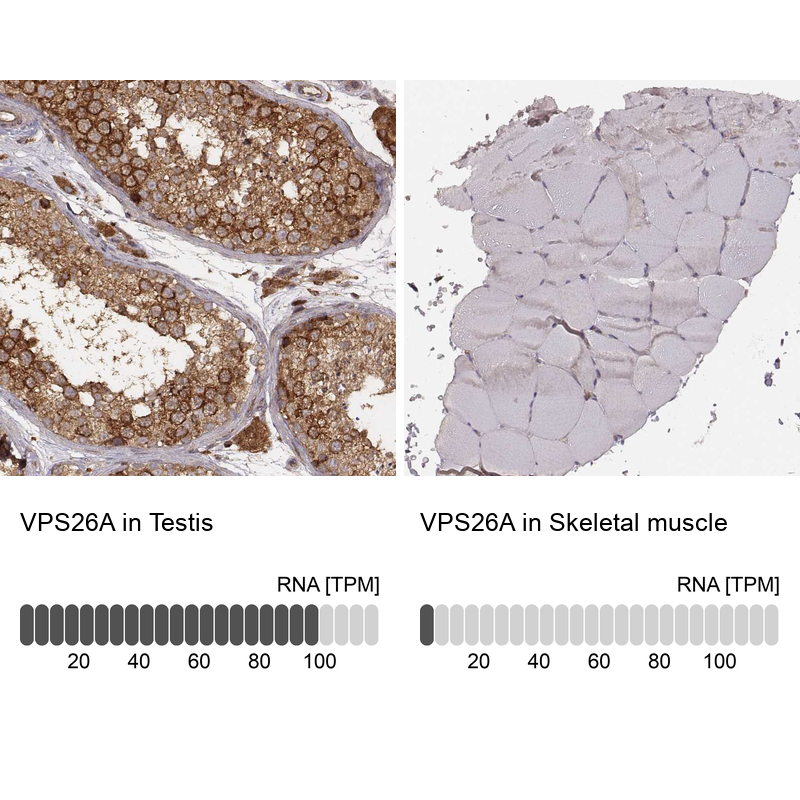
- Immunohistochemistry analysis in human testis and skeletal muscle tissues using AMAb90967 antibody. Corresponding VPS26A RNA-seq data are presented for the same tissues.
- Sample type
- Human
- Protocol
- Protocol