Explore
 Validate
 Learn
Learn Western blot
Antibody data
- Antibody Data
- Antigen structure
- References [6]
- Comments [0]
- Validations
- Western blot [3]
- Immunocytochemistry [1]
- Flow cytometry [1]
Submit
Validation data
Reference
Comment
Report error
- Product number
- PA1-972 - Provider product page

- Provider
- Invitrogen Antibodies
- Product name
- PSMB8 Polyclonal Antibody
- Antibody type
- Polyclonal
- Antigen
- Synthetic peptide
- Description
- PA1-972 detects Proteasome 20S LMP7 in western blot and immunofluorescence. PA1-972 can be used with blocking peptide PEP-132.
- Reactivity
- Human
- Host
- Rabbit
- Isotype
- IgG
- Vial size
- 100 µg
- Concentration
- 1 mg/mL
- Storage
- -20° C, Avoid Freeze/Thaw Cycles
Submitted references Evaluation of Immunoproteasome-Specific Proteolytic Activity Using Fluorogenic Peptide Substrates.
Human Lentiviral Gene Therapy Restores the Cellular Phenotype of Autosomal Recessive Complete IFN-γR1 Deficiency.
Active Protein Neddylation or Ubiquitylation Is Dispensable for Stress Granule Dynamics.
Glucosamine induces cell death via proteasome inhibition in human ALVA41 prostate cancer cell.
Inhibition of apoptosis in acute promyelocytic leukemia cells leads to increases in levels of oxidized protein and LMP2 immunoproteasome.
Neuronal induction of the immunoproteasome in Huntington's disease.
Kim S, Park SH, Choi WH, Lee MJ
Immune network 2022 Jun;22(3):e28
Immune network 2022 Jun;22(3):e28
Human Lentiviral Gene Therapy Restores the Cellular Phenotype of Autosomal Recessive Complete IFN-γR1 Deficiency.
Hahn K, Pollmann L, Nowak J, Nguyen AHH, Haake K, Neehus AL, Waqas SFH, Pessler F, Baumann U, Hetzel M, Casanova JL, Schulz A, Bustamante J, Ackermann M, Lachmann N
Molecular therapy. Methods & clinical development 2020 Jun 12;17:785-795
Molecular therapy. Methods & clinical development 2020 Jun 12;17:785-795
Active Protein Neddylation or Ubiquitylation Is Dispensable for Stress Granule Dynamics.
Markmiller S, Fulzele A, Higgins R, Leonard M, Yeo GW, Bennett EJ
Cell reports 2019 Apr 30;27(5):1356-1363.e3
Cell reports 2019 Apr 30;27(5):1356-1363.e3
Glucosamine induces cell death via proteasome inhibition in human ALVA41 prostate cancer cell.
Liu BQ, Meng X, Li C, Gao YY, Li N, Niu XF, Guan Y, Wang HQ
Experimental & molecular medicine 2011 Sep 30;43(9):487-93
Experimental & molecular medicine 2011 Sep 30;43(9):487-93
Inhibition of apoptosis in acute promyelocytic leukemia cells leads to increases in levels of oxidized protein and LMP2 immunoproteasome.
Khan MA, Oubrahim H, Stadtman ER
Proceedings of the National Academy of Sciences of the United States of America 2004 Aug 10;101(32):11560-5
Proceedings of the National Academy of Sciences of the United States of America 2004 Aug 10;101(32):11560-5
Neuronal induction of the immunoproteasome in Huntington's disease.
Díaz-Hernández M, Hernández F, Martín-Aparicio E, Gómez-Ramos P, Morán MA, Castaño JG, Ferrer I, Avila J, Lucas JJ
The Journal of neuroscience : the official journal of the Society for Neuroscience 2003 Dec 17;23(37):11653-61
The Journal of neuroscience : the official journal of the Society for Neuroscience 2003 Dec 17;23(37):11653-61
No comments: Submit comment
Supportive validation
- Submitted by
- Invitrogen Antibodies (provider)
- Main image

- Experimental details
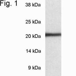
- Western blot of proteasome 20S LMP7 from HeLa cell extract using Product # PA1-972.

- Submitted by
- Invitrogen Antibodies (provider)
- Main image
- Experimental details
- Western blot of proteasome 20S LMP7 from HeLa cell extract using Product # PA1-972.

- Submitted by
- Invitrogen Antibodies (provider)
- Main image
- Experimental details
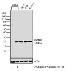
- Western blot analysis was performed on whole cell extracts (30 µglysate) of HeLa (Lane 1), HeLa treated with IFN gamma (100 ng/mL IFN gamma for 72h) (Lane 2), U-937 (Lane 3), Raji (Lane 4) and Ramos (Lane 5).The blots were probed with PSMB8 Rabbit polyclonal Antibody (Product # PA1-972, 2 µg/mL) and detected by chemiluminescence using Goat anti-Rabbit IgG (H+L) Superclonal™ Secondary Antibody, HRP conjugate (Product # A27036, 0.4 µg/mL, 1:2500 dilution). A 20 kDa band corresponding to PSMB9 was observed across the cell line tested and was enhanced upon IFN gamma treatment in HeLa cell line. Known quantity of protein samples were electrophoresed using Novex® NuPAGE® 4-12 % Bis-Tris gel (Product # NP0322BOX), XCell SureLock™ Electrophoresis System (Product # EI0002) and Novex® Sharp Pre-Stained Protein Standard (Product # LC5800). Resolved proteins were then transferred onto a nitrocellulose membrane with iBlot® 2 Dry Blotting System (Product # IB21001). The membrane was probed with the relevant primary and secondary Antibody following blocking with 5% skimmed milk. Chemiluminescent detection was performed using Pierce™ ECL Western Blotting Substrate (Product # 32106).

Supportive validation
- Submitted by
- Invitrogen Antibodies (provider)
- Main image
- Experimental details
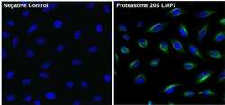
- Immunofluorescent analysis of Proteasome 20S LMP7 (green) in HeLa cells. The cells were fixed with 4% paraformaldehyde for 15 minutes, permeabilized with 0.1% Triton X-100 in TBS for 10 minutes, and blocked with 3% Blocker BSA (Product # 37525) in PBS for 15 minutes at room temperature. Cells were stained with or without Proteasome 20S LMP7 rabbit polyclonal antibody (Product # PA1-972), at a concentration of 5 µg/mL for 1 hour at room temperature, and then incubated with a Alexa Fluor® 488 Superclonal goat anti-rabbit IgG secondary antibody (Product # A27034) at a dilution of 1:1000 for 1 hour s at room temperature (both panels, green). Nuclei (both panels, blue) were stained with Hoechst 33342 dye (Product # 62249). Images were taken on a Thermo Scientific ToxInsight at 20X magnification.

Supportive validation
- Submitted by
- Invitrogen Antibodies (provider)
- Main image

- Experimental details
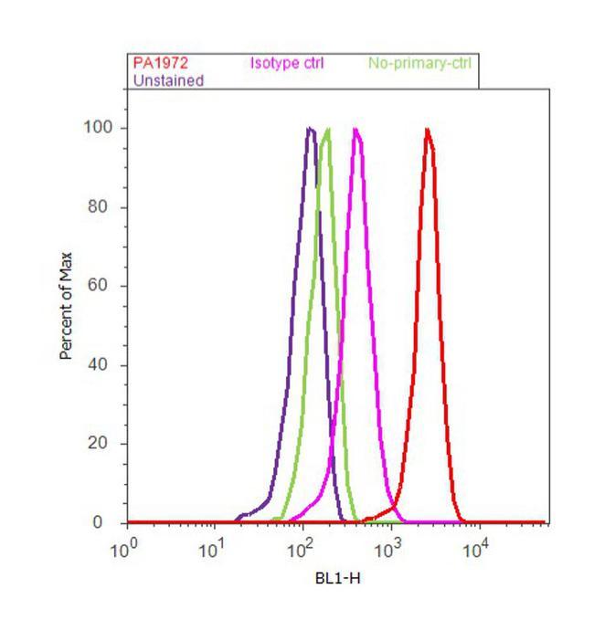
- Flow cytometry analysis of PSMB8 was done on SH-SY5Y cells. Cells were fixed with 70% ethanol for 10 minutes, permeabilized with 0.25% Triton™ X-100 for 20 minutes, and blocked with 5% BSA for 30 minutes at room temperature. Cells were labeled with PSMB8 Rabbit Polyclonal Antibody (PA1972, red histogram) or with rabbit isotype control (pink histogram) at 3-5 ug/million cells in 2.5% BSA. After incubation at room temperature for 2 hours, the cells were labeled with Alexa Fluor® 488 Goat Anti-Rabbit Secondary Antibody (A11008) at a dilution of 1:400 for 30 minutes at room temperature. The representative 10,000 cells were acquired and analyzed for each sample using an Attune® Acoustic Focusing Cytometer. The purple histogram represents unstained control cells and the green histogram represents no-primary-antibody control..