Explore
 Validate
 Learn
Learn Western blot
Antibody data
- Antibody Data
- Antigen structure
- References [0]
- Comments [0]
- Validations
- Western blot [1]
- Immunocytochemistry [1]
- Immunohistochemistry [4]
Submit
Validation data
Reference
Comment
Report error
- Product number
- UM800039CF - Provider product page

- Provider
- Invitrogen Antibodies
- Product name
- DOCK2 Monoclonal Antibody (UMAB142), UltraMAB™
- Antibody type
- Monoclonal
- Antigen
- Recombinant protein fragment
- Reactivity
- Human
- Host
- Mouse
- Isotype
- IgG
- Antibody clone number
- UMAB142
- Vial size
- 100 µg
- Concentration
- 1 mg/mL
- Storage
- -20° C, Avoid Freeze/Thaw Cycles
No comments: Submit comment
Supportive validation
- Submitted by
- Invitrogen Antibodies (provider)
- Main image
- Experimental details
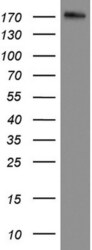
- Western blot analysis of RPMI8226 cell lysate (35 µg) by using anti-DOCK2 monoclonal antibody.

Supportive validation
- Submitted by
- Invitrogen Antibodies (provider)
- Main image

- Experimental details
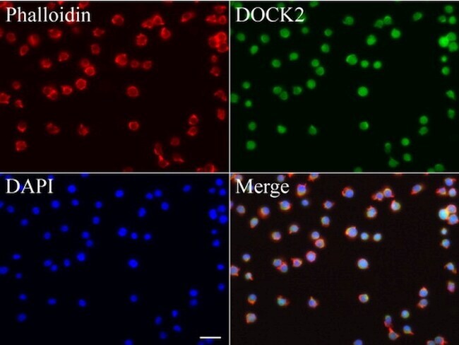
- Immunofluorescent staining of Jurkat cells using anti-DOCK2 mouse monoclonal antibody (UM800039, green, 1:100). Actin filaments were labeled with Alexa Fluor 594 Phalloidin (red), and nuclear with DAPI (blue). Scale bar, 20µm.
Supportive validation
- Submitted by
- Invitrogen Antibodies (provider)
- Main image

- Experimental details
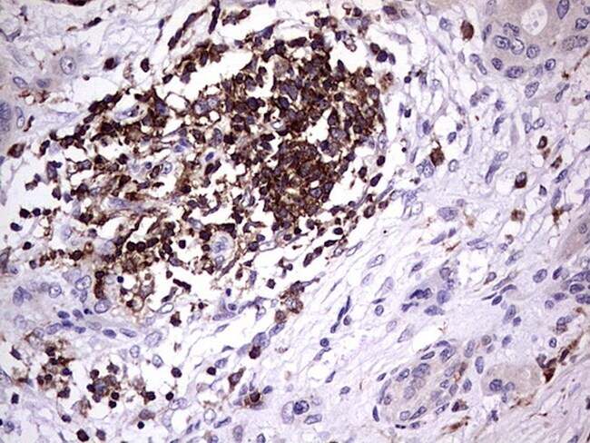
- Immunohistochemical staining of paraffin-embedded human lung cancer using anti-DOCK2 clone UMAB142 mouse monoclonal antibody at 1:200 dilution of mg/mL using Polink2 Broad HRP DAB for detection. UM800039 requires HIER with with citrate pH6.0 at 110°C for 3 min using pressure chamber/cooker. IHC staining shows tumor cells are negative however infiltrating lymphocytes strongly positive.
- Submitted by
- Invitrogen Antibodies (provider)
- Main image
- Experimental details
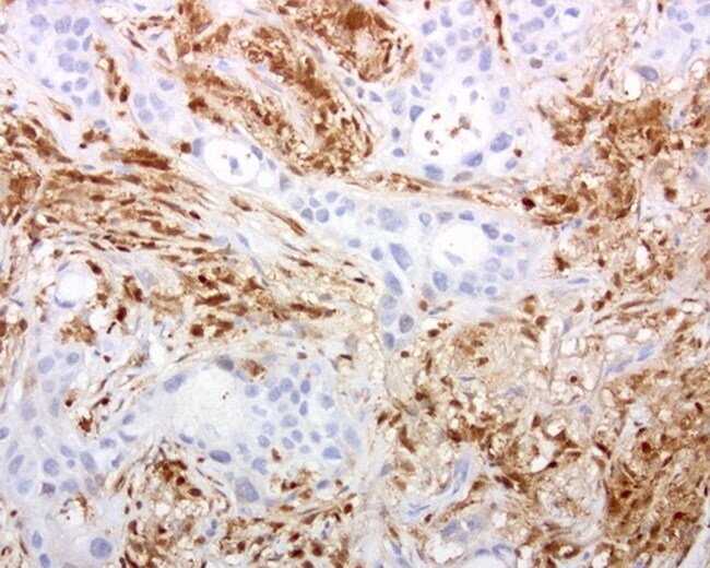
- Immunohistochemical staining of paraffin-embedded human melanoma using anti-DOCK2 clone UMAB142 mouse monoclonal antibody at 1:200 dilution of mg/mL using Polink2 Broad HRP DAB for detection. UM800039 requires HIER with with citrate pH6.0 at 110°C for 3 min using pressure chamber/cooker. IHC staining shows tumor cells are negative however infiltrating lymphocytes strongly positive.

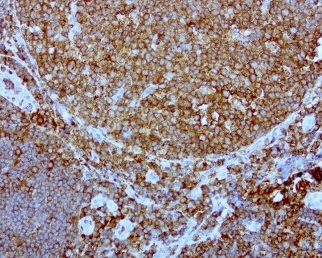
- Submitted by
- Invitrogen Antibodies (provider)
- Main image
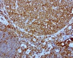
- Experimental details
- Immunohistochemical staining of paraffin-embedded human tonsil using anti-DOCK2 clone UMAB142 mouse monoclonal antibody at 1:200 dilution of mg/mL using Polink2 Broad HRP DAB for detection. UM800039 requires HIER with with citrate pH6.0 at 110°C for 3 min using pressure chamber/cooker. IHC staining shows germinal and nongerminal center with strong stain in the cytoplasm and membrane.
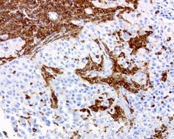
- Submitted by
- Invitrogen Antibodies (provider)
- Main image

- Experimental details
- Immunohistochemical staining of paraffin-embedded Carcinoma of Human pancreas tissue using anti-DOCK2 mouse monoclonal antibody. (UM800039; heat-induced epitope retrieval by 1 mM EDTA in 10mM Tris, pH8.5, 120°C for 3min)