 Explore
 Validate
 Learn
Learn Western blot
Antibody data
- Antibody Data
- Antigen structure
- References [0]
- Comments [0]
- Validations
- Western blot [1]
- Immunocytochemistry [2]
- Immunohistochemistry [2]
Submit
Validation data
Reference
Comment
Report error
- Product number
- PA1-24628 - Provider product page

- Provider
- Invitrogen Antibodies
- Product name
- OPRM1 Polyclonal Antibody
- Antibody type
- Polyclonal
- Antigen
- Synthetic peptide
- Description
- PA1-24628 is expected to cross react with bovine (93% conserved), guinea pig (100% conserved), Macaque monkey (100% conserved), and porcine (100% conserved) due to sequence homology.
- Concentration
- Conc. Not Determined
No comments: Submit comment
Supportive validation
- Submitted by
- Invitrogen Antibodies (provider)
- Main image

- Experimental details
- Western blot was performed using OPRM1 Polyclonal Antibody (Product # PA1-24628) and ~53kDa band corresponding to OPRM1 was observed across cell lines and tissue extracts tested except in Mouse Kidney and Mouse Heart which are reported to be negative. An uncharacterized band (*) at ~110, ~15 kDa was observed in the Rat Brain and Mouse Lungs respectively. Whole cell extracts (30 µg lysate) of (Fig. a) SH-SY5Y (Lane 1), SK-N-AS (Lane 2), IMR-32 (Lane 3), U-2 OS (Lane 4), MCF7 (Lane 5), Neuro-2a (Lane 6), A549 (Lane 7), tissue extracts (30ug lysate) of Mouse Testis (Lane 8), Mouse Brain (Lane 9), Rat Brain (Lane 10) and Mouse Lungs (Lane 11); (Fig. b) tissue extracts (30ug lysate) of Mouse Brain (Lane 1), Rat Brain (Lane 2), Mouse Testis (Lane 3), Rat Testis (Lane 4), Mouse Lungs (Lane 5), Mouse Kidney (Lane 6) and Mouse Heart (Lane 7) were electrophoresed using NuPAGE® 10% Bis-Tris gel (Product # NP0302BOX). Resolved proteins were then transferred onto a nitrocellulose membrane (Product # IB23001) by iBlot® 2 Dry Blotting System (Product # IB21001).The blot was probed with the primary antibody (1:1000 dilution) and detected by chemiluminescence with Goat anti-Rabbit IgG (H+L) Superclonal™ Recombinant Secondary Antibody, HRP (Product # A27036, 1:4000 dilution) using the iBright FL 1000 (Product # A32752). Chemiluminescent detection was performed using Novex® ECL Chemiluminescent Substrate Reagent Kit (Product # WP20005)..

Supportive validation
- Submitted by
- Invitrogen Antibodies (provider)
- Main image

- Experimental details
- Immunofluorescent analysis of mu Opioid Receptor using a mu Opioid Receptor polyclonal antibody (Product # PA1-24628). The image shows the presence of a subpopulation of spinal lamina II neurons that contained both µ opioid receptor (green) and TRPV1 (red) immunoreactivity in a vehicle (A)- and a RTX (B)-treated rats. Co-localization of the µ opioid receptor and TRPV1 immunoreactivity is indicated in yellow when 2 images are digitally merged. All images are single confocal optical sections. Scale bar: 20 µm.

- Submitted by
- Invitrogen Antibodies (provider)
- Main image

- Experimental details
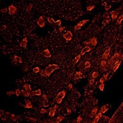
- Immunofluorescent analysis of mu Opioid Receptor in rat DRG using a mu Opioid Receptor polyclonal antibody (Product # PA1-24628) at a dilution of 1:100. Cells were incubated at 4ºC overnight and detected using an anti-Rabbit Rhodamine Red secondary antibody at a dilution of 1:200, incubated at room temperature for 1 hour.

Supportive validation
- Submitted by
- Invitrogen Antibodies (provider)
- Main image
- Experimental details
- Immunohistochemistry analysis of rat DRG tissue using OPRM1 Polyclonal Antibody (Product # PA1-24628). Dilution: 1:100.

- Submitted by
- Invitrogen Antibodies (provider)
- Main image
- Experimental details
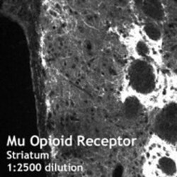
- Immunohistochemistry (Frozen) analysis of striatum using (Product # PA1-24628) OPRM1 Polyclonal Antibody.
