Explore
 Validate
 Learn
Learn Western blot
Antibody data
- Antibody Data
- Antigen structure
- References [1]
- Comments [0]
- Validations
- Western blot [2]
- Immunohistochemistry [1]
Submit
Validation data
Reference
Comment
Report error
- Product number
- AF4818 - Provider product page

- Provider
- Novus Biologicals
- Product name
- Sheep Polyclonal VSIG1 Antibody
- Antibody type
- Polyclonal
- Description
- Immunogen affinity purified. Detects human VSIG1 in direct ELISAs and Western blots. In direct ELISAs and Western blots, approximately 5% cross-reactivity with recombinant human (rh) VSIG2, rhVSIG3, rhVSIG4, and recombinant mouse VSIG1 is observed.
- Reactivity
- Human
- Host
- Sheep
- Conjugate
- Unconjugated
- Isotype
- IgG
- Vial size
- 100 ug
- Concentration
- LYOPH
- Storage
- Use a manual defrost freezer and avoid repeated freeze-thaw cycles. 12 months from date of receipt, -20 to -70 degreesC as supplied. 1 month, 2 to 8 degreesC under sterile conditions after reconstitution. 6 months, -20 to -70 degreesC under sterile conditions after reconstitution.
Submitted references RNA sequencing of sessile serrated colon polyps identifies differentially expressed genes and immunohistochemical markers.
Delker DA, McGettigan BM, Kanth P, Pop S, Neklason DW, Bronner MP, Burt RW, Hagedorn CH
PloS one 2014;9(2):e88367
PloS one 2014;9(2):e88367
No comments: Submit comment
Supportive validation
- Submitted by
- Novus Biologicals (provider)
- Main image
- Experimental details
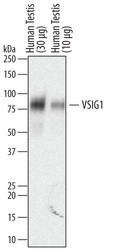
- Detection of Human VSIG1 by Western Blot. Western blot shows lysates of human testis tissue. PVDF membrane was probed with 0.2 µg/mL of Sheep Anti-Human VSIG1 Antigen Affinity-purified Polyclonal Antibody (Catalog # AF4818) followed by HRP-conjugated Anti-Sheep IgG Secondary Antibody (Catalog # HAF016). A specific band was detected for VSIG1 at approximately 80 kDa (as indicated). This experiment was conducted under reducing conditions and using Immunoblot Buffer Group 1.

- Submitted by
- Novus Biologicals (provider)
- Main image

- Experimental details
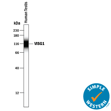
- Detection of Human VSIG1 by Simple WesternTM. Simple Western lane view shows lysates of human testis tissue, loaded at 0.2 mg/mL. A specific band was detected for VSIG1 at approximately 115 kDa (as indicated) using 10 µg/mL of Sheep Anti-Human VSIG1 Antigen Affinity-purified Polyclonal Antibody (Catalog # AF4818) followed by 1:50 dilution of HRP-conjugated Anti-Sheep IgG Secondary Antibody (Catalog # HAF016). This experiment was conducted under reducing conditions and using the 12-230 kDa separation system.
Supportive validation
- Submitted by
- Novus Biologicals (provider)
- Main image

- Experimental details
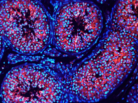
- VSIG1 in Human Testis. VSIG1 was detected in immersion fixed frozen sections of human testis using 10 µg/mL Sheep Anti-Human VSIG1 Antigen Affinity-purified Polyclonal Antibody (Catalog # AF4818) overnight at 4 °C. Tissue was stained with the NorthernLights™ 557-conjugated Anti-Sheep IgG Secondary Antibody (red; Catalog # NL010) and counterstained with DAPI (blue). View our protocol for Fluorescent IHC Staining of Frozen Tissue Sections.