Explore
 Validate
 Learn
Learn Western blot
Antibody data
- Antibody Data
- Antigen structure
- References [0]
- Comments [0]
- Validations
- Western blot [1]
- Immunohistochemistry [1]
- Flow cytometry [1]
Submit
Validation data
Reference
Comment
Report error
- Product number
- MAB48181-100 - Provider product page

- Provider
- R&D Systems
- Product name
- Human VSIG1 Antibody
- Antibody type
- Monoclonal
- Description
- Protein A or G purified from cell culture supernatant. Detects human VSIG-1 in direct ELISAs.
- Reactivity
- Human
- Host
- Rabbit
- Conjugate
- Unconjugated
- Antigen sequence
Q86XK7- Isotype
- IgG
- Antibody clone number
- 1314D
- Vial size
- 100 ug
- Storage
- Use a manual defrost freezer and avoid repeated freeze-thaw cycles. 12 months from date of receipt, -20 to -70 °C as supplied. 1 month, 2 to 8 °C under sterile conditions after reconstitution. 6 months, -20 to -70 °C under sterile conditions after reconstitution.
No comments: Submit comment
Supportive validation
- Submitted by
- R&D Systems (provider)
- Main image
- Experimental details
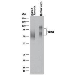
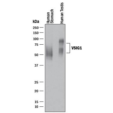
- Detection of Human VSIG1 by Western Blot. Western blot shows lysates of human stomach tissue and human testis tissue. PVDF membrane was probed with 0.5 µg/mL of Rabbit Anti-Human VSIG1 Monoclonal Antibody (Catalog # MAB48181) followed by HRP-conjugated Anti-Rabbit IgG Secondary Antibody (Catalog # HAF008). Specific bands were detected for VSIG1 at approximately 55-75 kDa (as indicated). This experiment was conducted under reducing conditions and using Immunoblot Buffer Group 1.
Supportive validation
- Submitted by
- R&D Systems (provider)
- Main image
- Experimental details
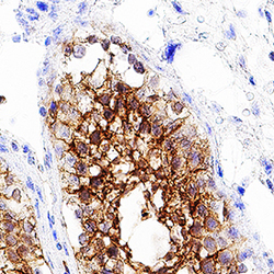
- VSIG1 in Human Testis. VSIG1 was detected in immersion fixed paraffin-embedded sections of human testis using Rabbit Anti-Human VSIG1 Monoclonal Antibody (Catalog # MAB48181) at 1 µg/mL for 1 hour at room temperature followed by incubation with the Anti-Rabbit IgG VisUCyte™ HRP Polymer Antibody (Catalog # VC003). Before incubation with the primary antibody, tissue was subjected to heat-induced epitope retrieval using Antigen Retrieval Reagent-Basic (Catalog # CTS013). Tissue was stained using DAB (brown) and counterstained with hematoxylin (blue). Specific staining was localized to cell membrane and cytoplasm. View our protocol for IHC Staining with VisUCyte HRP Polymer Detection Reagents.

Supportive validation
- Submitted by
- R&D Systems (provider)
- Main image
- Experimental details
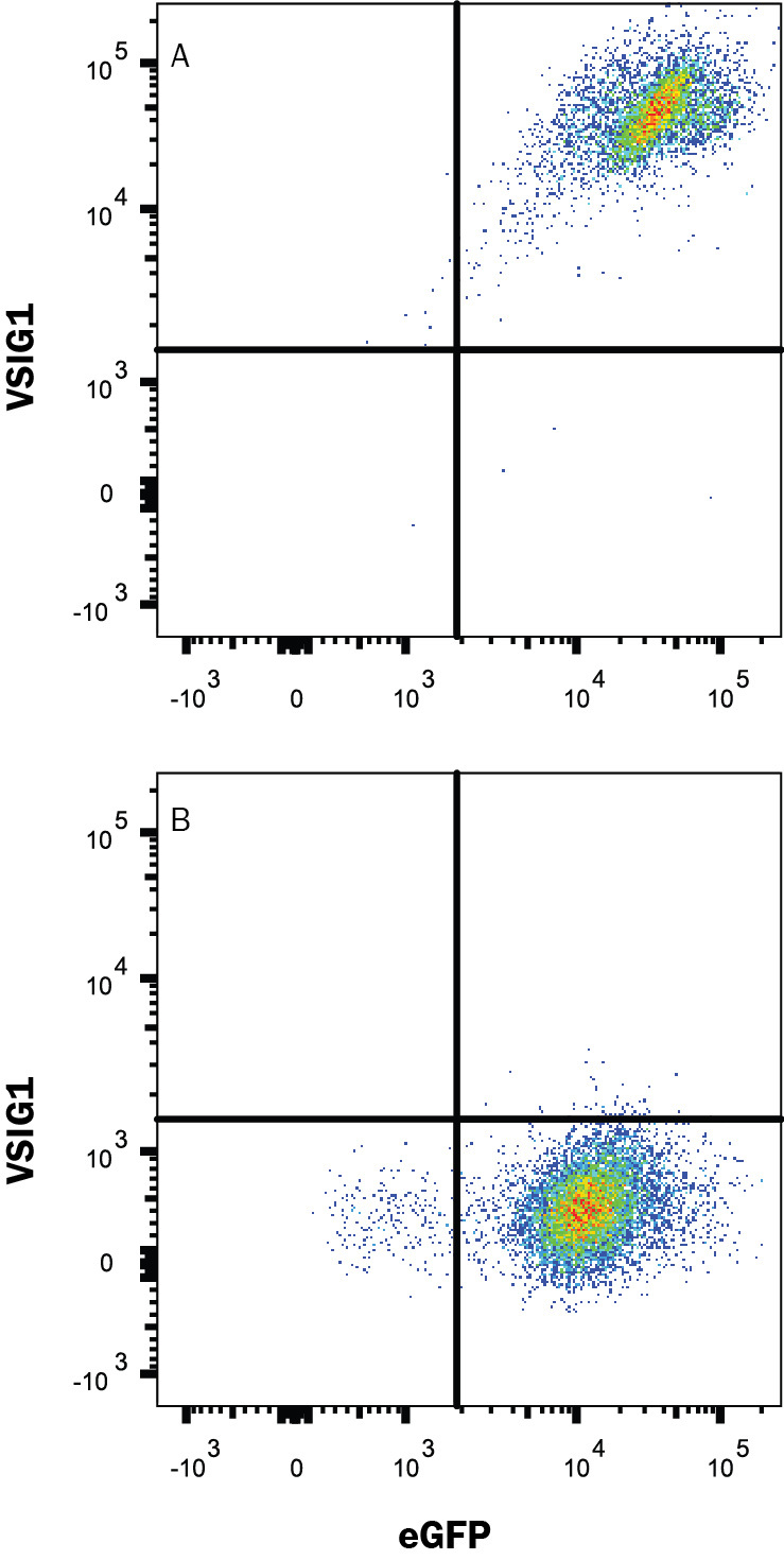
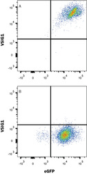
- Detection of VSIG1 in HEK293 Human Cell Line Transfected with Human VSIG1 and eGFP by Flow Cytometry. HEK293 human embryonic kidney cell line transfected with either (A) human VSIG1 or (B) irrelevant transfectants and eGFP was stained with Rabbit Anti-Human VSIG1 Monoclonal Antibody (Catalog # MAB48181) followed by Allophycocyanin-conjugated Anti-Rabbit IgG Secondary Antibody (Catalog # F0111). Quadrant markers were set based on control antibody staining (Catalog # MAB1050). View our protocol for Staining Membrane-associated Proteins.