Explore
 Validate
 Learn
Learn Western blot
 Immunohistochemistry
Antibody data
- Antibody Data
- Antigen structure
- References [1]
- Comments [0]
- Validations
- Immunohistochemistry [1]
Submit
Validation data
Reference
Comment
Report error
- Product number
- AF1188 - Provider product page

- Provider
- R&D Systems
- Product name
- Rat GABAB R2 N-Terminus Antibody
- Antibody type
- Polyclonal
- Description
- Antigen Affinity-purified. Detects rat GABAB R2 N-Terminus in direct ELISAs and Western blots.
- Reactivity
- Rat
- Host
- Goat
- Conjugate
- Unconjugated
- Antigen sequence
O88871- Isotype
- IgG
- Vial size
- 100 ug
- Concentration
- LYOPH
- Storage
- Use a manual defrost freezer and avoid repeated freeze-thaw cycles. 12 months from date of receipt, -20 to -70 °C as supplied. 1 month, 2 to 8 °C under sterile conditions after reconstitution. 6 months, -20 to -70 °C under sterile conditions after reconstitution.
Submitted references Modular composition and dynamics of native GABAB receptors identified by high-resolution proteomics.
Schwenk J, Pérez-Garci E, Schneider A, Kollewe A, Gauthier-Kemper A, Fritzius T, Raveh A, Dinamarca MC, Hanuschkin A, Bildl W, Klingauf J, Gassmann M, Schulte U, Bettler B, Fakler B
Nature neuroscience 2016 Feb;19(2):233-42
Nature neuroscience 2016 Feb;19(2):233-42
No comments: Submit comment
Supportive validation
- Submitted by
- R&D Systems (provider)
- Main image
- Experimental details
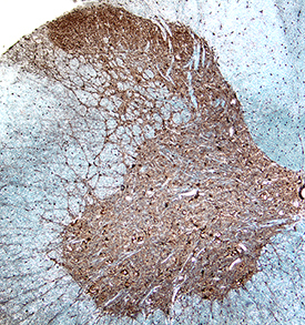
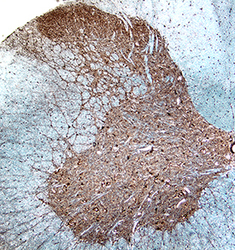
- GABAB R2 in Rat Spinal Cord. GABAB R2 was detected in perfusion fixed frozen sections of rat spinal cord using Goat Anti-Rat GABAB R2 N-Terminus Antigen Affinity-purified Polyclonal Antibody (Catalog # AF1188) at 15 µg/mL for 1 hour at room temperature followed by incubation with the Anti-Goat IgG VisUCyte™ HRP Polymer Antibody (Catalog # VC004). Tissue was stained using DAB (brown) and counterstained with hematoxylin (blue). Specific staining was localized to dorsal horn. View our protocol for IHC Staining with VisUCyte HRP Polymer Detection Reagents.