Explore
 Validate
 Learn
Learn Immunocytochemistry
Antibody data
- Antibody Data
- Antigen structure
- References [0]
- Comments [0]
- Validations
- Immunocytochemistry [1]
- Immunohistochemistry [1]
Submit
Validation data
Reference
Comment
Report error
- Product number
- 703357 - Provider product page

- Provider
- Invitrogen Antibodies
- Product name
- GABBR2 Recombinant Rabbit Monoclonal Antibody (19H3L12)
- Antibody type
- Monoclonal
- Antigen
- Synthetic peptide
- Reactivity
- Human, Rat
- Host
- Rabbit
- Isotype
- IgG
- Antibody clone number
- 19H3L12
- Vial size
- 100 µg
- Concentration
- 0.5 mg/mL
- Storage
- Store at 4°C short term. For long term storage, store at -20°C, avoiding freeze/thaw cycles.
No comments: Submit comment
Supportive validation
- Submitted by
- Invitrogen Antibodies (provider)
- Main image
- Experimental details
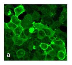
- For immunofluorescence analysis, HEK-293 cells were stably transfected with human GABBR2. After 24hr of transfection the cells were fixed and permeabilized for detection of GABBR2 using Anti-GABBR2 Recombinant Rabbit Monoclonal Antibody (Product # 703357, 1 µg/mL, 1:500 dilution) and labeled with Goat anti-Rabbit IgG (H+L) Superclonal™ Secondary Antibody, Alexa Fluor® 488 conjugate. Panel a) shows representative cells that were stained for detection of membrane localization of the GABBR2 on the transfected cells. The images were captured at 40X magnification.

Supportive validation
- Submitted by
- Invitrogen Antibodies (provider)
- Main image

- Experimental details
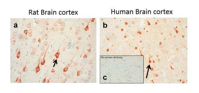
- Sections of Rat and Human Brain cortex were dewaxed, microwaved in citric acid and incubated with Anti-GABBR2 Recombinant Rabbit Monoclonal Antibody (Product # 703357, 1 µg/mL, 1:500 dilution). Sections were then sequentially treated with biotinylated Anti-rabbit IgG and AB solution. Sections were further developed in AEC and lightly counterstained with hematoxylin. Panel a & b) shows the specific localization of GABBR2 in neuronal stomata and dendrites in both Rat and Human brain cortex respectively. Panel c) shows the no primary antibody control.