 Explore
 Validate
 Learn
LearnAF6739
antibody from Novus Biologicals
Targeting: CELSR2
ADGRC2, CDHF10, EGFL2, Flamingo1, KIAA0279, MEGF3
 Western blot
Antibody data
- Antibody Data
- Antigen structure
- References [1]
- Comments [0]
- Validations
- Western blot [1]
- Immunohistochemistry [1]
- Flow cytometry [2]
Submit
Validation data
Reference
Comment
Report error
- Product number
- AF6739 - Provider product page

- Provider
- Novus Biologicals
- Product name
- Goat Polyclonal CELSR2 Antibody
- Antibody type
- Polyclonal
- Description
- Immunogen affinity purified. Detects human CELSR2 in direct ELISAs and Western blots. In direct ELISAs, less than 5% cross-reactivity with recombinant human (rh) CELSR1 and rhCELSR3 is observed.
- Reactivity
- Human, Mouse
- Host
- Goat
- Isotype
- IgG
- Vial size
- 100 ug
- Concentration
- LYOPH
- Storage
- Use a manual defrost freezer and avoid repeated freeze-thaw cycles. 12 months from date of receipt, -20 to -70 degreesC as supplied. 1 month, 2 to 8 degreesC under sterile conditions after reconstitution. 6 months, -20 to -70 degreesC under sterile conditions after reconstitution.
Submitted references SNX27 Deletion Causes Hydrocephalus by Impairing Ependymal Cell Differentiation and Ciliogenesis.
Wang X, Zhou Y, Wang J, Tseng IC, Huang T, Zhao Y, Zheng Q, Gao Y, Luo H, Zhang X, Bu G, Hong W, Xu H
The Journal of neuroscience : the official journal of the Society for Neuroscience 2016 Dec 14;36(50):12586-12597
The Journal of neuroscience : the official journal of the Society for Neuroscience 2016 Dec 14;36(50):12586-12597
No comments: Submit comment
Supportive validation
- Submitted by
- Novus Biologicals (provider)
- Main image

- Experimental details
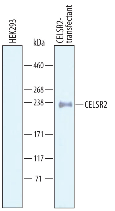
- Detection of CELSR2 by Western Blot. Western blot shows lysates of HEK293 human embryonic kidney cell line either mock transfected or transfected with human CELSR2. PVDF Membrane was probed with 1 µg/mL of Goat Anti-Human CELSR2 Antigen Affinity-purified Polyclonal Antibody (Catalog # AF6739) followed by HRP-conjugated Anti-Goat IgG Secondary Antibody (Catalog # HAF019). A specific band was detected for CELSR2 at approximately 240 kDa (as indicated). This experiment was conducted under reducing conditions and using Immunoblot Buffer Group 8.
Supportive validation
- Submitted by
- Novus Biologicals (provider)
- Main image

- Experimental details
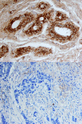
- CELSR2 in Human Breast. CELSR2 was detected in immersion fixed paraffin-embedded sections of human breast using Goat Anti-Human CELSR2 Antigen Affinity-purified Polyclonal Antibody (Catalog # AF6739) at 10 µg/mL overnight at 4 °C. Tissue was stained using the Anti-Goat HRP-DAB Cell & Tissue Staining Kit (brown; Catalog # CTS008) and counterstained with hematoxylin (blue). Lower panel shows a lack of labeling when primary antibodies are omitted and tissue is stained only with secondary antibody followed by incubation with detection reagents. Specific staining was localized to ductal epithelium. View our protocol for Chromogenic IHC Staining of Paraffin-embedded Tissue Sections.
Supportive validation
- Submitted by
- Novus Biologicals (provider)
- Main image
- Experimental details
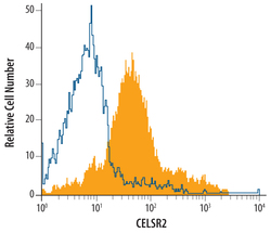
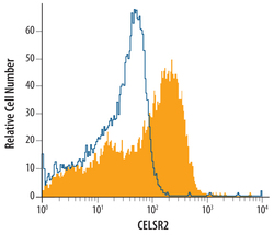
- Detection of CELSR2 in SH-SY5Y Human Cell Line by Flow Cytometry. SH-SY5Y human neuroblastoma cell line was stained with Goat Anti-Human CELSR2 Antigen Affinity-purified Polyclonal Antibody (Catalog # AF6739, filled histogram) or isotype control antibody (Catalog # AB-108-C, open histogram), followed by Allophycocyanin-conjugated Anti-Goat IgG Secondary Antibody (Catalog # F0108).
- Submitted by
- Novus Biologicals (provider)
- Main image
- Experimental details
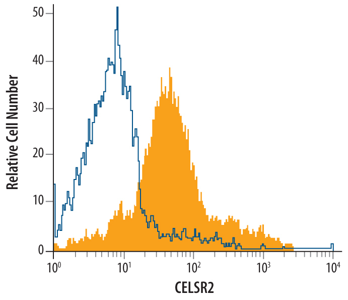
- Detection of CELSR2 in bEnd.3 Mouse Cell Line by Flow Cytometry. bEnd.3 mouse endothelioma cell line was stained with Goat Anti-Human CELSR2 Antigen Affinity-purified Polyclonal Antibody (Catalog # AF6739, filled histogram) or isotype control antibody (Catalog # AB-108-C, open histogram), followed by Allophycocyanin-conjugated Anti-Goat IgG Secondary Antibody (Catalog # F0108).
