Explore
 Validate
 Learn
Learn Western blot
 Immunocytochemistry
Antibody data
- Antibody Data
- Antigen structure
- References [3]
- Comments [0]
- Validations
- Immunocytochemistry [4]
Submit
Validation data
Reference
Comment
Report error
- Product number
- BML-SA610-0200 - Provider product page

- Provider
- Enzo Life Sciences
- Product name
- mGluR1 (extracellular), pAb
- Antibody type
- Polyclonal
- Antigen
- Synthetic peptide
- Description
- Lyophilized from PBS, pH 7.4, 1% BSA and 0.025% sodium azide.
- Vial size
- 200 µl
Submitted references Metabotropic glutamate receptors (mGluR5) activate transient receptor potential canonical channels to improve the regularity of the respiratory rhythm generated by the pre-Bötzinger complex in mice.
Deleterious effects of amyloid beta oligomers acting as an extracellular scaffold for mGluR5.
Disassembly of shank and homer synaptic clusters is driven by soluble beta-amyloid(1-40) through divergent NMDAR-dependent signalling pathways.
Ben-Mabrouk F, Amos LB, Tryba AK
The European journal of neuroscience 2012 Jun;35(11):1725-37
The European journal of neuroscience 2012 Jun;35(11):1725-37
Deleterious effects of amyloid beta oligomers acting as an extracellular scaffold for mGluR5.
Renner M, Lacor PN, Velasco PT, Xu J, Contractor A, Klein WL, Triller A
Neuron 2010 Jun 10;66(5):739-54
Neuron 2010 Jun 10;66(5):739-54
Disassembly of shank and homer synaptic clusters is driven by soluble beta-amyloid(1-40) through divergent NMDAR-dependent signalling pathways.
Roselli F, Hutzler P, Wegerich Y, Livrea P, Almeida OF
PloS one 2009 Jun 23;4(6):e6011
PloS one 2009 Jun 23;4(6):e6011
No comments: Submit comment
Supportive validation
- Submitted by
- Enzo Life Sciences (provider)
- Main image

- Experimental details
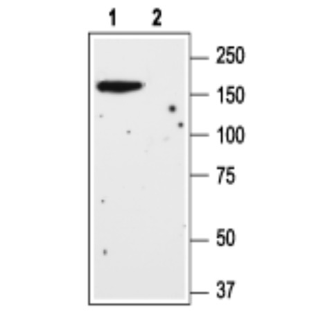
- Western blot analysis of rat brain lysate: 1. Anti-mGluR1 (extracellular) antibody (Cat# BML-SA610), (1:200). 2. Anti-mGluR1 (extracellular) antibody, preincubated with the control peptide antigen.
- Submitted by
- Enzo Life Sciences (provider)
- Main image
- Experimental details
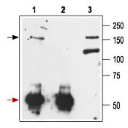
- Immunoprecipitation of rat brain lysate:. 1. Cell lysate + protein A beads + Anti-mGluR1 (extracellular) antibody (Prod. No. BML-SA610). 2. Cell lysate + protein A beads + pre-immune rabbit serum. 3. Cell lysate. Black arrow indicates the mGluR1 protein (glycosylated form) while the red arrow shows the IgG heavy chain. Immunoblot was performed with the Anti-mGluR1 (extracellular) antibody.

- Submitted by
- Enzo Life Sciences (provider)
- Main image
- Experimental details
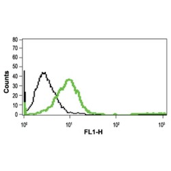
- Indirect flow cytometry analysis in live intact Jurkat cells:. Black: Unstained cells + goat-anti-rabbit-FITC. Green: Cells + Anti-mGluR1 (extracellular) antibody (Cat# BML-SA610), (10 µg) + goat-anti-rabbit-FITC.

- Submitted by
- Enzo Life Sciences (provider)
- Main image
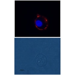
- Experimental details
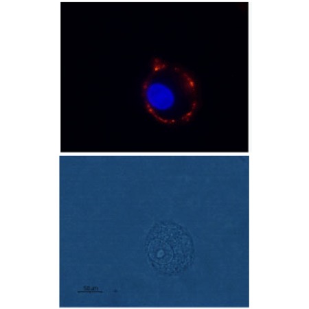
- Expression of mGluR1 in rat C6 glioma cells: Immunocytochemical staining of live intact rat C6 glioma cells using Anti-mGluR1 (extracellular) antibody (Cat# BML-SA610), (1:100), followed by goat-anti-rabbit-AlexaFluor-555 secondary antibody (red). Nuclei were stained with Hoechst 33342 (blue).