Explore
 Validate
 Learn
Learn Immunohistochemistry
Antibody data
- Antibody Data
- Antigen structure
- References [2]
- Comments [0]
- Validations
- Immunohistochemistry [1]
Submit
Validation data
Reference
Comment
Report error
- Product number
- MAB45141 - Provider product page

- Provider
- R&D Systems
- Product name
- Human mGluR5 Antibody
- Antibody type
- Monoclonal
- Description
- Protein A or G purified from hybridoma culture supernatant. Detects human mGLUR in direct ELISAs and Western blots.
- Reactivity
- Human
- Host
- Mouse
- Conjugate
- Unconjugated
- Antigen sequence
P41594- Isotype
- IgG
- Antibody clone number
- 464823
- Vial size
- 100 ug
- Concentration
- LYOPH
- Storage
- Use a manual defrost freezer and avoid repeated freeze-thaw cycles. 12 months from date of receipt, -20 to -70 °C as supplied. 1 month, 2 to 8 °C under sterile conditions after reconstitution. 6 months, -20 to -70 °C under sterile conditions after reconstitution.
Submitted references The role of metabotropic glutamate receptor 5 on the stromal cell-derived factor-1/CXCR4 system in oral cancer.
Delayed mGluR5 activation limits neuroinflammation and neurodegeneration after traumatic brain injury.
Kuribayashi N, Uchida D, Kinouchi M, Takamaru N, Tamatani T, Nagai H, Miyamoto Y
PloS one 2013;8(11):e80773
PloS one 2013;8(11):e80773
Delayed mGluR5 activation limits neuroinflammation and neurodegeneration after traumatic brain injury.
Byrnes KR, Loane DJ, Stoica BA, Zhang J, Faden AI
Journal of neuroinflammation 2012 Feb 28;9:43
Journal of neuroinflammation 2012 Feb 28;9:43
No comments: Submit comment
Supportive validation
- Submitted by
- R&D Systems (provider)
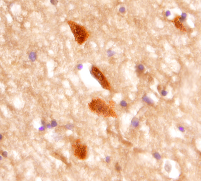
- Main image

- Experimental details
- mGluR5 in Human Brain. mGluR5 was detected in immersion fixed paraffin-embedded sections of human brain (cerebellum) using 25 µg/mL Mouse Anti-Human mGluR5 Monoclonal Antibody (Catalog # MAB45141) overnight at 4 °C. Tissue was stained with the Anti-Mouse HRP-DAB Cell & Tissue Staining Kit (brown; Catalog # CTS002) and counterstained with hematoxylin (blue). View our protocol for Chromogenic IHC Staining of Paraffin-embedded Tissue Sections.