Explore
 Validate
 Learn
LearnMA1-16622
antibody from Invitrogen Antibodies
Targeting: EP300
KAT3B, p300
 Western blot Immunocytochemistry
 Immunoprecipitation Flow cytometry Chromatin Immunoprecipitation Other assay
Antibody data
- Antibody Data
- Antigen structure
- References [2]
- Comments [0]
- Validations
- Western blot [2]
- Immunocytochemistry [8]
- Flow cytometry [4]
- Other assay [1]
Submit
Validation data
Reference
Comment
Report error
- Product number
- MA1-16622 - Provider product page

- Provider
- Invitrogen Antibodies
- Product name
- p300 Monoclonal Antibody (RW105)
- Antibody type
- Monoclonal
- Antigen
- Other
- Description
- Suggested positive control: HeLa nuclear extract.
- Reactivity
- Human, Mouse, Rat
- Host
- Mouse
- Isotype
- IgG
- Antibody clone number
- RW105
- Vial size
- 100 μL
- Concentration
- 1 mg/mL
- Storage
- -20°C, Avoid Freeze/Thaw Cycles
Submitted references Tumor necrosis factor-alpha inhibitors suppress CCL2 chemokine in monocytes via epigenetic modification.
Reactive oxygen species, AMP-activated protein kinase, and the transcription cofactor p300 regulate α-tubulin acetyltransferase-1 (αTAT-1/MEC-17)-dependent microtubule hyperacetylation during cell stress.
Lin YC, Lin YC, Huang MY, Kuo PL, Wu CC, Lee MS, Hsieh CC, Kuo HF, Kuo CH, Tsai WC, Hung CH
Molecular immunology 2017 Mar;83:82-91
Molecular immunology 2017 Mar;83:82-91
Reactive oxygen species, AMP-activated protein kinase, and the transcription cofactor p300 regulate α-tubulin acetyltransferase-1 (αTAT-1/MEC-17)-dependent microtubule hyperacetylation during cell stress.
Mackeh R, Lorin S, Ratier A, Mejdoubi-Charef N, Baillet A, Bruneel A, Hamaï A, Codogno P, Poüs C, Perdiz D
The Journal of biological chemistry 2014 Apr 25;289(17):11816-11828
The Journal of biological chemistry 2014 Apr 25;289(17):11816-11828
No comments: Submit comment
Supportive validation
- Submitted by
- Invitrogen Antibodies (provider)
- Main image
- Experimental details
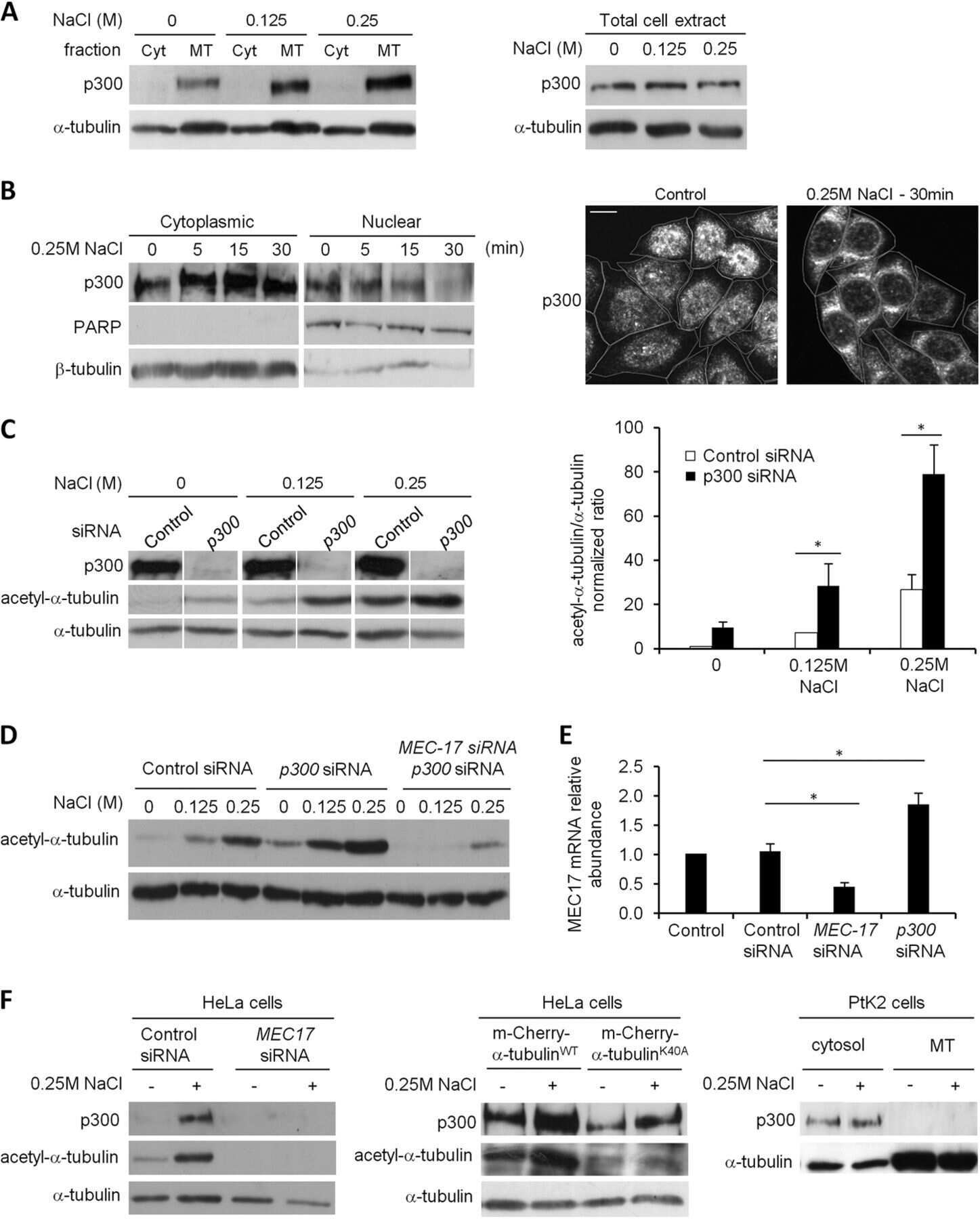
- Western blot analysis of p300 in HeLa nuclear extract. Samples were incubated in p300 monoclonal antibody (Product # MA1-16622 using a dilution of 1:250. ECL: 20 minute exposure.

- Submitted by
- Invitrogen Antibodies (provider)
- Main image
- Experimental details
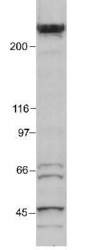
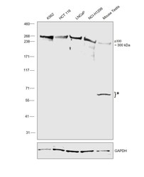
- Western blot was performed using Anti-p300 Monoclonal Antibody (RW105) (Product # MA1-16622) and a 300 kDa band corresponding to p300 was observed across cell lines and tissue tested. Nuclear enriched extracts (40 µg lysate) of K-562 (Lane 1), HCT 116 (Lane 2), LNCaP (Lane 3), H1299 (Lane 4) and Mouse Testis (Lane 5) were electrophoresed using NuPAGE™ 3-8% Tris-Acetate Protein Gel (Product # EA0378BOX). Resolved proteins were then transferred onto a nitrocellulose membrane (Product # IB23001) by iBlot® 2 Dry Blotting System (Product # IB21001) and then equilibrated with 20% ethanol. The blot was probed with the primary antibody (1:1000 dilution) and detected by chemiluminescence with Goat anti-Mouse IgG (H+L) Superclonal™ Recombinant Secondary Antibody, HRP (Product # A28177,1:20000 dilution) using the iBright™ FL1500 Imaging System (Product # A44115). Chemiluminescent detection was performed using SuperSignal™ West Pico PLUS Chemiluminescent Substrate (Product # 34580).
Supportive validation
- Submitted by
- Invitrogen Antibodies (provider)
- Main image

- Experimental details
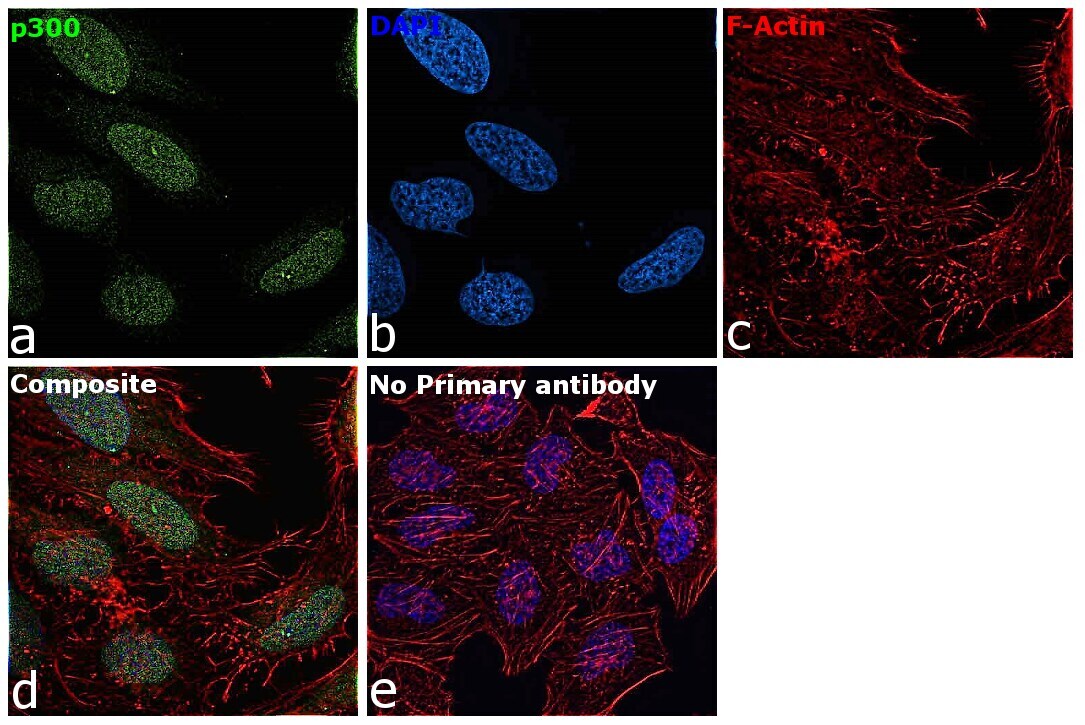
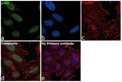
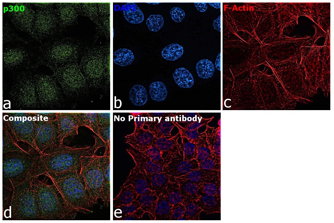
- Immunofluorescence analysis of p300 Monoclonal Antibody (RW105) was performed using 70% confluent log phase HeLa cells. The cells were fixed with 4% paraformaldehyde for 10 minutes, permeabilized with 0.1% Triton™ X-100 for 15 minutes, and blocked with 2% BSA for 45 minutes at room temperature. The cells were labeled with p300 Monoclonal Antibody (RW105) (Product # MA1-16622) at 1:100 dilution in 0.1% BSA, incubated at 4 degree celsius overnight and then labeled with Donkey anti-Mouse IgG (H+L) Highly Cross-Adsorbed Secondary Antibody, Alexa Fluor Plus 488 (Product # A32766), (1:2000 dilution), for 45 minutes at room temperature (Panel a: Green). Nuclei (Panel b:Blue) were stained with ProLong™ Diamond Antifade Mountant with DAPI (Product # P36962). F-actin (Panel c: Red) was stained with Rhodamine Phalloidin (Product # R415, 1:300 dilution). Panel d represents the merged image showing nuclear as well as cytoplasmic localization. Panel e represents control cells with no primary antibody to assess background. The images were captured at 60X magnification.
- Submitted by
- Invitrogen Antibodies (provider)
- Main image
- Experimental details
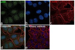
- Immunofluorescence analysis of p300 Monoclonal Antibody (RW105) was performed using 70% confluent log phase HCT 116 cells. The cells were fixed with 4% paraformaldehyde for 10 minutes, permeabilized with 0.1% Triton™ X-100 for 15 minutes, and blocked with 2% BSA for 45 minutes at room temperature. The cells were labeled with p300 Monoclonal Antibody (RW105) (Product # MA1-16622) at 1:100 dilution in 0.1% BSA, incubated at 4 degree celsius overnight and then labeled with Donkey anti-Mouse IgG (H+L) Highly Cross-Adsorbed Secondary Antibody, Alexa Fluor Plus 488 (Product # A32766), (1:2,000 dilution), for 45 minutes at room temperature (Panel a: Green). Nuclei (Panel b:Blue) were stained with ProLong™ Diamond Antifade Mountant with DAPI (Product # P36962). F-actin (Panel c: Red) was stained with Rhodamine Phalloidin (Product # R415, 1:300 dilution). Panel d represents the merged image showing nuclear localization. Panel e represents control cells with no primary antibody to assess background. The images were captured at 60X magnification.

- Submitted by
- Invitrogen Antibodies (provider)
- Main image

- Experimental details
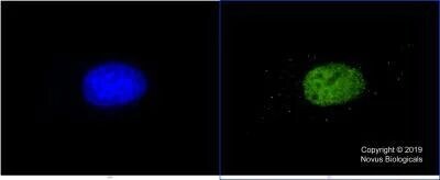
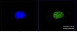
- Immunocytochemistry analysis of p300 in HeLa cells fixed in 4% paraformaldehyde for 10 min and permeabilized in 0.05% Triton X-100 in PBS for 5 minutes. Samples were incubated in p300 monoclonal antibody (Product # MA1-16622) using a dilution of 5 µg/mL for 60 minutes at room temperature followed by anti-mouse DyLight 488 (Green) with a dilution of 1:1000 dilution for 60 minutes. Nuclei were counterstained with DAPI (Blue). Cells were imaged using a 100X objective.
- Submitted by
- Invitrogen Antibodies (provider)
- Main image

- Experimental details
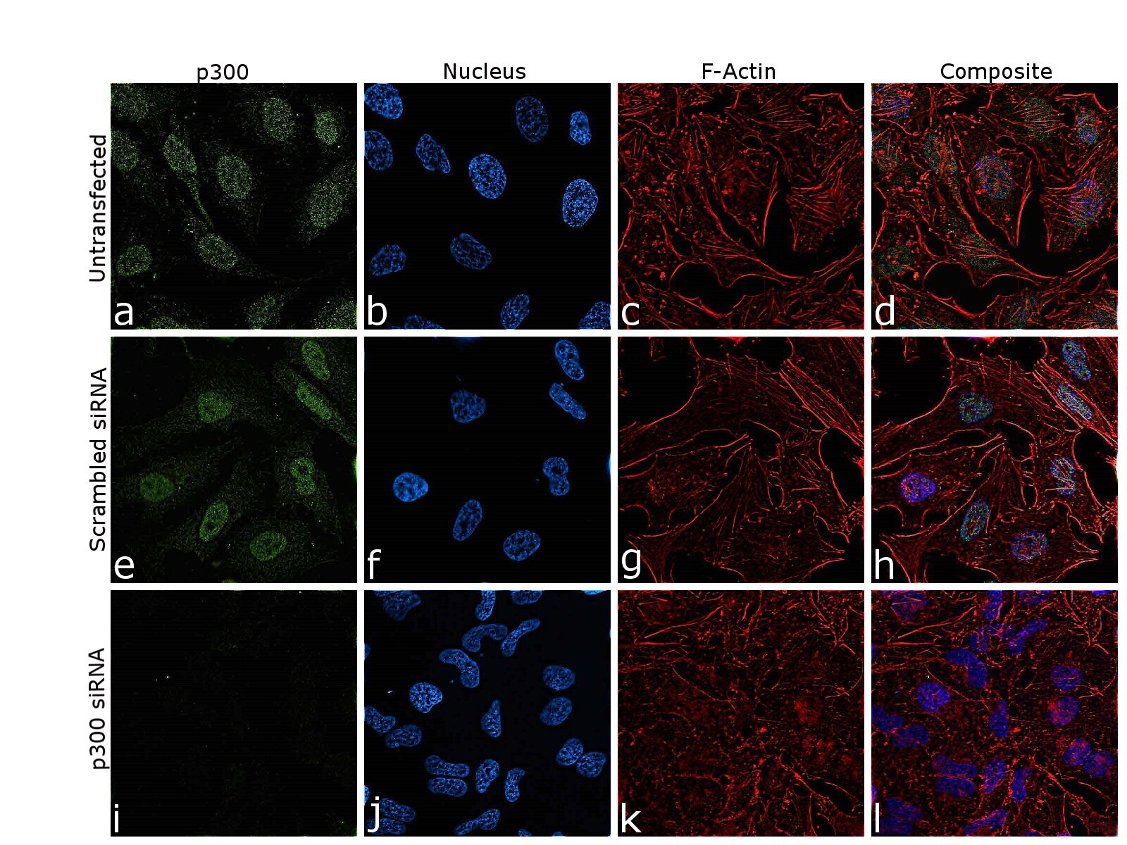
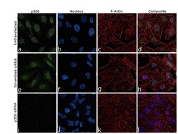
- Knockdown of p300 was achieved by transfecting HeLa cells with p300 Monoclonal Antibody (RW105) specific siRNA (Silencer® select Product # S4696, S4697). Immunofluorescence analysis was performed on untransfected HeLa cells (panel a-d), transfected with non-specific scrambled siRNA (panels e-h) and transfected with p300 specific siRNA (panel i-l). Cells were fixed, permeabilized, and labelled with p300 Monoclonal Antibody (RW105) (Product # MA1-16622, 1:100 dilution) followed by Donkey anti-Mouse IgG (H+L) Highly Cross-Adsorbed Secondary Antibody, Alexa Fluor Plus 488 (Product # A32766), (1:2000 dilution). Nuclei (blue) were stained using ProLong™ Diamond Antifade Mountant with DAPI (Product # P36962), and Rhodamine Phalloidin (Product # R415, 1:300 dilution) was used for cytoskeletal F-actin (Red) staining. reduction in signal of specific signal was observed upon siRNA mediated knockdown (panel i-l) confirming specificity of the antibody to p300 (Green). The Images were captured at 60X magnification.
- Submitted by
- Invitrogen Antibodies (provider)
- Main image
- Experimental details
- Immunocytochemistry analysis of p300 in HeLa cells fixed in 4% paraformaldehyde for 10 min and permeabilized in 0.05% Triton X-100 in PBS for 5 minutes. Samples were incubated in p300 monoclonal antibody (Product # MA1-16622) using a dilution of 5 µg/mL for 60 minutes at room temperature followed by anti-mouse DyLight 488 (Green) with a dilution of 1:1000 dilution for 60 minutes. Nuclei were counterstained with DAPI (Blue). Cells were imaged using a 100X objective.

- Submitted by
- Invitrogen Antibodies (provider)
- Main image
- Experimental details
- Knockdown of p300 was achieved by transfecting HeLa cells with p300 Monoclonal Antibody (RW105) specific siRNA (Silencer® select Product # S4696, S4697). Immunofluorescence analysis was performed on untransfected HeLa cells (panel a-d), transfected with non-specific scrambled siRNA (panels e-h) and transfected with p300 specific siRNA (panel i-l). Cells were fixed, permeabilized, and labelled with p300 Monoclonal Antibody (RW105) (Product # MA1-16622, 1:100 dilution) followed by Donkey anti-Mouse IgG (H+L) Highly Cross-Adsorbed Secondary Antibody, Alexa Fluor Plus 488 (Product # A32766), (1:2000 dilution). Nuclei (blue) were stained using ProLong™ Diamond Antifade Mountant with DAPI (Product # P36962), and Rhodamine Phalloidin (Product # R415, 1:300 dilution) was used for cytoskeletal F-actin (Red) staining. reduction in signal of specific signal was observed upon siRNA mediated knockdown (panel i-l) confirming specificity of the antibody to p300 (Green). The Images were captured at 60X magnification.

- Submitted by
- Invitrogen Antibodies (provider)
- Main image
- Experimental details
- Immunofluorescence analysis of p300 Monoclonal Antibody (RW105) was performed using 70% confluent log phase HeLa cells. The cells were fixed with 4% paraformaldehyde for 10 minutes, permeabilized with 0.1% Triton™ X-100 for 15 minutes, and blocked with 2% BSA for 45 minutes at room temperature. The cells were labeled with p300 Monoclonal Antibody (RW105) (Product # MA1-16622) at 1:100 dilution in 0.1% BSA, incubated at 4 degree celsius overnight and then labeled with Donkey anti-Mouse IgG (H+L) Highly Cross-Adsorbed Secondary Antibody, Alexa Fluor Plus 488 (Product # A32766), (1:2000 dilution), for 45 minutes at room temperature (Panel a: Green). Nuclei (Panel b:Blue) were stained with ProLong™ Diamond Antifade Mountant with DAPI (Product # P36962). F-actin (Panel c: Red) was stained with Rhodamine Phalloidin (Product # R415, 1:300 dilution). Panel d represents the merged image showing nuclear as well as cytoplasmic localization. Panel e represents control cells with no primary antibody to assess background. The images were captured at 60X magnification.

- Submitted by
- Invitrogen Antibodies (provider)
- Main image

- Experimental details
- Immunofluorescence analysis of p300 Monoclonal Antibody (RW105) was performed using 70% confluent log phase HCT 116 cells. The cells were fixed with 4% paraformaldehyde for 10 minutes, permeabilized with 0.1% Triton™ X-100 for 15 minutes, and blocked with 2% BSA for 45 minutes at room temperature. The cells were labeled with p300 Monoclonal Antibody (RW105) (Product # MA1-16622) at 1:100 dilution in 0.1% BSA, incubated at 4 degree celsius overnight and then labeled with Donkey anti-Mouse IgG (H+L) Highly Cross-Adsorbed Secondary Antibody, Alexa Fluor Plus 488 (Product # A32766), (1:2,000 dilution), for 45 minutes at room temperature (Panel a: Green). Nuclei (Panel b:Blue) were stained with ProLong™ Diamond Antifade Mountant with DAPI (Product # P36962). F-actin (Panel c: Red) was stained with Rhodamine Phalloidin (Product # R415, 1:300 dilution). Panel d represents the merged image showing nuclear localization. Panel e represents control cells with no primary antibody to assess background. The images were captured at 60X magnification.
Supportive validation
- Submitted by
- Invitrogen Antibodies (provider)
- Main image

- Experimental details
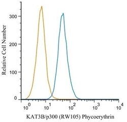
- Flow cytometry of p300 in RAW 246.7 cells (blue) and a matched isotype control (orange). Samples were incubated in p300 monoclonal antibody (Product # MA1-16622) using a dilution of 2 µg/mL for 30 minutes at room temperature. Cells were fixed with 4% PFA and then permeablized with 0.1% saponin. Both antibodies were conjugated to Phycoerythrin.

- Submitted by
- Invitrogen Antibodies (provider)
- Main image

- Experimental details
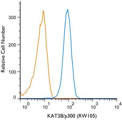
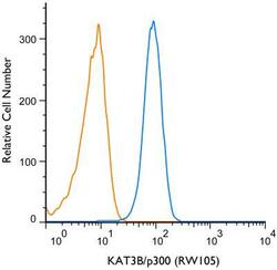
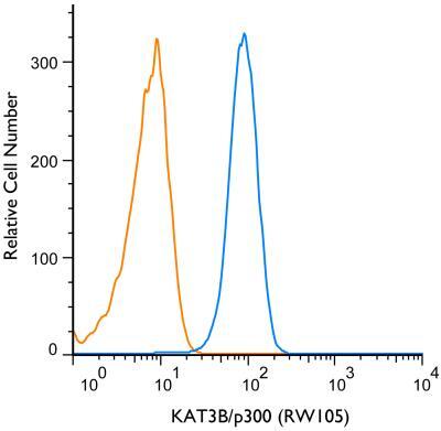
- Flow cytometry of p300 in THP-1 cells. Samples were incubated in p300 monoclonal antibody (Product # MA1-16622) and a matched isotype control using a dilution of 1 µg/mL for 30 minutes at room temperature followed by mouse F(ab)2 IgG (H+L) APC-conjugated secondary antibody. Antibody (blue) and a matched isotype control (orange). Cells were fixed with 4% PFA and permeabilized with 0.1% Saponin.
- Submitted by
- Invitrogen Antibodies (provider)
- Main image
- Experimental details
- Flow cytometry of p300 in RAW 246.7 cells (blue) and a matched isotype control (orange). Samples were incubated in p300 monoclonal antibody (Product # MA1-16622) using a dilution of 2 µg/mL for 30 minutes at room temperature. Cells were fixed with 4% PFA and then permeablized with 0.1% saponin. Both antibodies were conjugated to Phycoerythrin.

- Submitted by
- Invitrogen Antibodies (provider)
- Main image
- Experimental details
- Flow cytometry of p300 in THP-1 cells. Samples were incubated in p300 monoclonal antibody (Product # MA1-16622) and a matched isotype control using a dilution of 1 µg/mL for 30 minutes at room temperature followed by mouse F(ab)2 IgG (H+L) APC-conjugated secondary antibody. Antibody (blue) and a matched isotype control (orange). Cells were fixed with 4% PFA and permeabilized with 0.1% Saponin.
Supportive validation
- Submitted by
- Invitrogen Antibodies (provider)
- Main image

- Experimental details
- NULL