Explore
 Validate
 Learn
Learn Western blot
 Immunocytochemistry
Antibody data
- Antibody Data
- Antigen structure
- References [0]
- Comments [0]
- Validations
- Immunocytochemistry [4]
Submit
Validation data
Reference
Comment
Report error
- Product number
- BML-SA609-0050 - Provider product page

- Provider
- Enzo Life Sciences
- Proper citation
- Enzo Life Sciences Cat#BML-SA609-0050, RRID:AB_2050764
- Product name
- AT2 receptor (extracellular), pAb
- Antibody type
- Polyclonal
- Antigen
- Synthetic peptide
- Description
- Lyophilized from PBS, pH 7.4, 1% BSA and 0.025% sodium azide.
- Vial size
- 50 µl
No comments: Submit comment
Supportive validation
- Submitted by
- Enzo Life Sciences (provider)
- Main image
- Experimental details
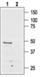
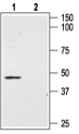
- Image 1: Western blot of rat brain membranes: Lane 1.ÊPAb to AT2 Receptor (Extracellular)Ê(Prod. No. BML-SA609), 1:500. Lane 2. PAb to AT2 Receptor (Extracellular), preincubated with the control peptide antigen.

- Submitted by
- Enzo Life Sciences (provider)
- Main image
- Experimental details
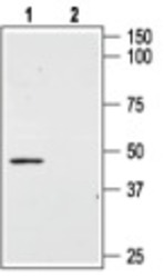
- Image 2: Western blot of mouse MS1 endothelial cells: Lane 1. PAb to AT2 Receptor (Extracellular) (Prod. No. BML-SA609), 1:400. Lane 2. PAb to AT2 Receptor (Extracellular), preincubated with the control peptide antigen.
- Submitted by
- Enzo Life Sciences (provider)
- Main image
- Experimental details
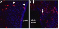
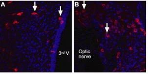
- Image 3: Staining of angiotensin II receptor type-2 (AT2) withÊPAb to AT2 Receptor (Extracellular)Ê(Prod. No. BML-SA609) in rat brain sections. AT2 receptor is stained red, a Nissl counterstain is in blue. A, AT2 receptor expressing neurons are scattered in the paraventricular nucleus of the hypothalamus (arrows), in the vicinity of the 3rd ventricle (3rd V). B, AT2 receptor expressing neurons in the supraoptic nucleus (arrows), adjacent to the optic nerve.
- Submitted by
- Enzo Life Sciences (provider)
- Main image

- Experimental details
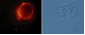
- Image 4: Immunocytochemical staining of live intact rat GH3 pituitary cells withÊPAb to AT2 Receptor (Extracellular)Ê(Prod. No. BML-SA609) (1:100), followed by goat-anti-rabbit-AlexaFluor-555 secondary antibody.