Explore
 Validate
 Learn
LearnNBP2-31374
antibody from Novus Biologicals
Targeting: CCR5
CC-CKR-5, CD195, CKR-5, CKR5, CMKBR5, IDDM22
 Western blot
Antibody data
- Antibody Data
- Antigen structure
- References [2]
- Comments [0]
- Validations
- Western blot [3]
- Immunohistochemistry [4]
Submit
Validation data
Reference
Comment
Report error
- Product number
- NBP2-31374 - Provider product page

- Provider
- Novus Biologicals
- Product name
- Rabbit Polyclonal CCR5 Antibody
- Antibody type
- Polyclonal
- Description
- Protein A purified.
- Reactivity
- Human, Mouse, Simian
- Host
- Rabbit
- Isotype
- IgG
- Vial size
- 0.1 mg
- Concentration
- 1.0 mg/ml
- Storage
- Store at 4C short term. Aliquot and store at -20C long term. Avoid freeze-thaw cycles.
Submitted references Methamphetamine abuse affects gene expression in brain-derived microglia of SIV-infected macaques to enhance inflammation and promote virus targets.
JNK-1 deficiency limits macrophage-mediated antigen-induced arthritis.
Najera JA, Bustamante EA, Bortell N, Morsey B, Fox HS, Ravasi T, Marcondes MC
BMC immunology 2016 Apr 23;17(1):7
BMC immunology 2016 Apr 23;17(1):7
JNK-1 deficiency limits macrophage-mediated antigen-induced arthritis.
Guma M, Ronacher LM, Firestein GS, Karin M, Corr M
Arthritis and rheumatism 2011 Jun;63(6):1603-12
Arthritis and rheumatism 2011 Jun;63(6):1603-12
No comments: Submit comment
Supportive validation
- Submitted by
- Novus Biologicals (provider)
- Main image
- Experimental details
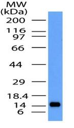
- Western Blot: CCR5 Antibody [NBP2-31374] - Detection of CCR5 partial recombinant protein by using CCR5 antibody at a concentration of 0.5 ug/mL.

- Submitted by
- Novus Biologicals (provider)
- Main image

- Experimental details
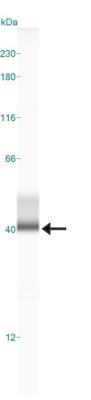
- Simple Western: CCR5 Antibody [NBP2-31374] - Lane view shows a specific band for CCR5 in 0.5 mg/mL of MCF-7 lysate. This experiment was performed under reducing conditions using the 12-230 kDa separation system.
- Submitted by
- Novus Biologicals (provider)
- Main image

- Experimental details
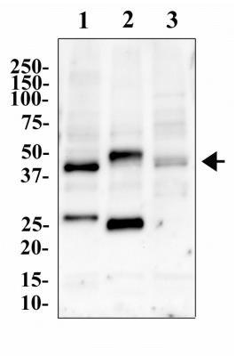
- Western Blot: CCR5 Antibody [NBP2-31374] - Image of anti-CCR5 antibody. Whole cell protein from Daudi (1), K562 (2) and THP-1 (3) cell lines were separated by SDS-PAGE and protein transfered to PVDF. The membrane was probed with anti-CCR5 antibody at 2 ug/mL and detected with an anti-rabbit-HRP secondary antibody and chemiluminescence. CCR5 is shown at 40 kDa.
Supportive validation
- Submitted by
- Novus Biologicals (provider)
- Main image

- Experimental details
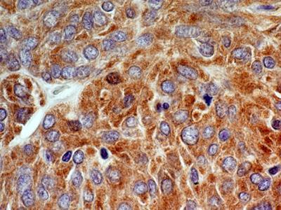
- Immunohistochemistry-Paraffin: CCR5 Antibody [NBP2-31374] - Analysis of CCR5 protein in a section of malignant stromal tumor of small bowel from human using CCR5 antibody at a concentration of 5 ug/mL.
- Submitted by
- Novus Biologicals (provider)
- Main image
- Experimental details
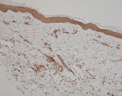
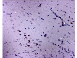
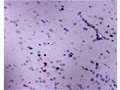
- Immunohistochemistry-Paraffin: CCR5 Antibody [NBP2-31374] - Staining in a Rhesus macaque brain. IHC-P image submitted by a verified customer review.
- Submitted by
- Novus Biologicals (provider)
- Main image

- Experimental details
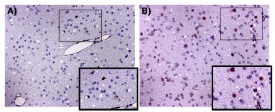
- Immunohistochemistry-Paraffin: CCR5 Antibody [NBP2-31374] - Detection of CCR5 and virus on brain tissue. CCR5 expression was analyzed using Immunohistochemistry on the layer III of frontal cortex from macaques that were uninfected (A) and untreated controls, and Meth-only treated (B). Pictures were at 16x magnification. Sites of interest (rectangles) were further magnified. Image collected and cropped by CiteAb from the following publication (http://bmcimmunol.biomedcentral.com/articles/10.1186/s12865-016-0145-0), licensed under a CC-BY licence.
- Submitted by
- Novus Biologicals (provider)
- Main image
- Experimental details
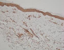
- Immunohistochemistry-Paraffin: CCR5 Antibody [NBP2-31374] - Human skin section. IHC-P image submitted by a verified customer review.