Explore
 Validate
 Learn
Learn Western blot
 Immunocytochemistry
Antibody data
- Antibody Data
- Antigen structure
- References [0]
- Comments [0]
- Validations
- Immunocytochemistry [11]
- Immunohistochemistry [1]
- Flow cytometry [6]
Submit
Validation data
Reference
Comment
Report error
- Product number
- PA5-79857 - Provider product page

- Provider
- Invitrogen Antibodies
- Product name
- MYPT1 Polyclonal Antibody
- Antibody type
- Polyclonal
- Antigen
- Synthetic peptide
- Description
- Reconstitute with 0.2 mL of distilled water to yield a concentration of 500 µg/mL. Positive Control - WB: human HELA whole cell, human Jurkat whole cell, human HEK293 whole cell, monkey COS-7 whole cell, human Raji whole cell, human K562 whole cell, human CACO-2 whole cell, human HEPG2 whole cell, rat brain tissue, rat lung tissue, rat liver tissue, rat C6 whole cell, mouse brain tissue, mouse lung tissue, mouse liver tissue, mouse NIH/3T3 whole cell. IHC: Human Glioma tissue. ICC/IF: SiHa cell, U251 cell, U251 cell, U251 cell, U251 cell. Flow: Hela cell, SiHa cell, U251 cell.
- Reactivity
- Human, Mouse, Rat
- Host
- Rabbit
- Isotype
- IgG
- Vial size
- 100 μg
- Concentration
- 500 μg/mL
- Storage
- -20°C
No comments: Submit comment
Supportive validation
- Submitted by
- Invitrogen Antibodies (provider)
- Main image
- Experimental details
- Immunofluorescent analysis of MYPT1 on SiHa cells. Antigen retrieval was performed using citrate buffer (pH6, epitope retrieval solution) for 20 mins. Sample was blocked using 10% goat serum, incubated with MYPT1 polyclonal antibody (Product# PA5-79857) with a dilution of 2 µg/mL (overnight at 4°C). Development was performed using Streptavidin-Biotin-Complex (SABC) with DAB chromogen method.
- Submitted by
- Invitrogen Antibodies (provider)
- Main image
- Experimental details
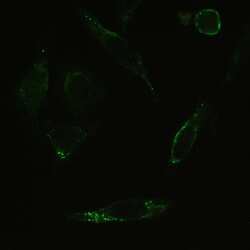
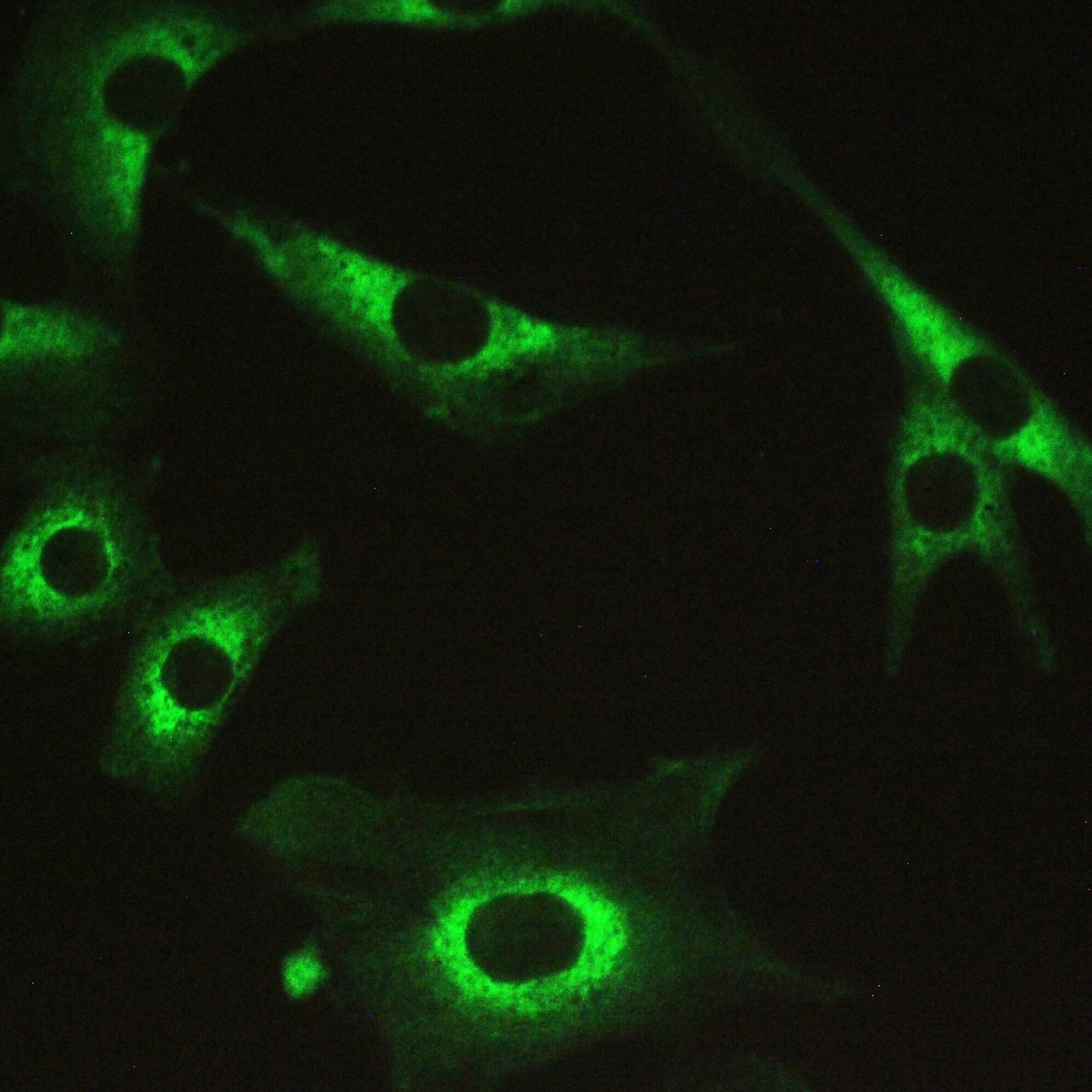
- Immunofluorescent analysis of MYPT1 on U251 cells. Antigen retrieval was performed using citrate buffer (pH6, epitope retrieval solution) for 20 mins. Sample was blocked using 10% goat serum, incubated with MYPT1 polyclonal antibody (Product# PA5-79857) with a dilution of 2 µg/mL (overnight at 4°C). Development was performed using Streptavidin-Biotin-Complex (SABC) with DAB chromogen method.
- Submitted by
- Invitrogen Antibodies (provider)
- Main image
- Experimental details
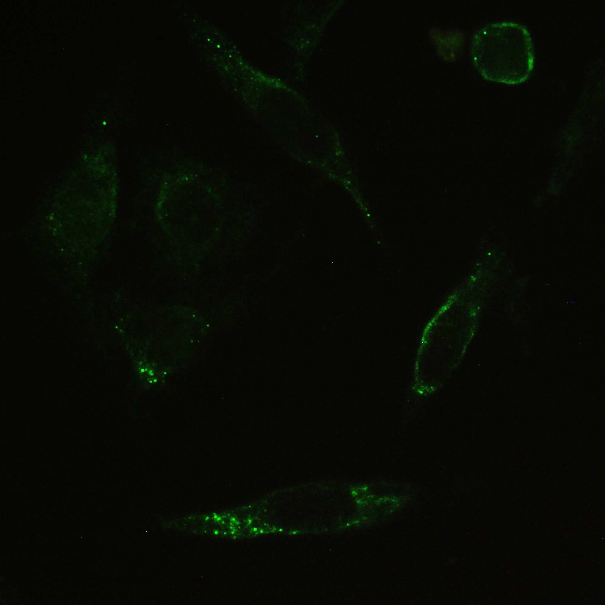
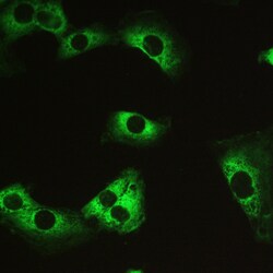
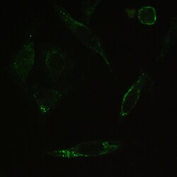
- Immunofluorescent analysis of MYPT1 on U251 cells. Antigen retrieval was performed using citrate buffer (pH6, epitope retrieval solution) for 20 mins. Sample was blocked using 10% goat serum, incubated with MYPT1 polyclonal antibody (Product# PA5-79857) with a dilution of 2 µg/mL (overnight at 4°C). Development was performed using Streptavidin-Biotin-Complex (SABC) with DAB chromogen method.

- Submitted by
- Invitrogen Antibodies (provider)
- Main image
- Experimental details
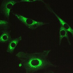
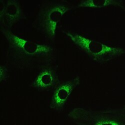
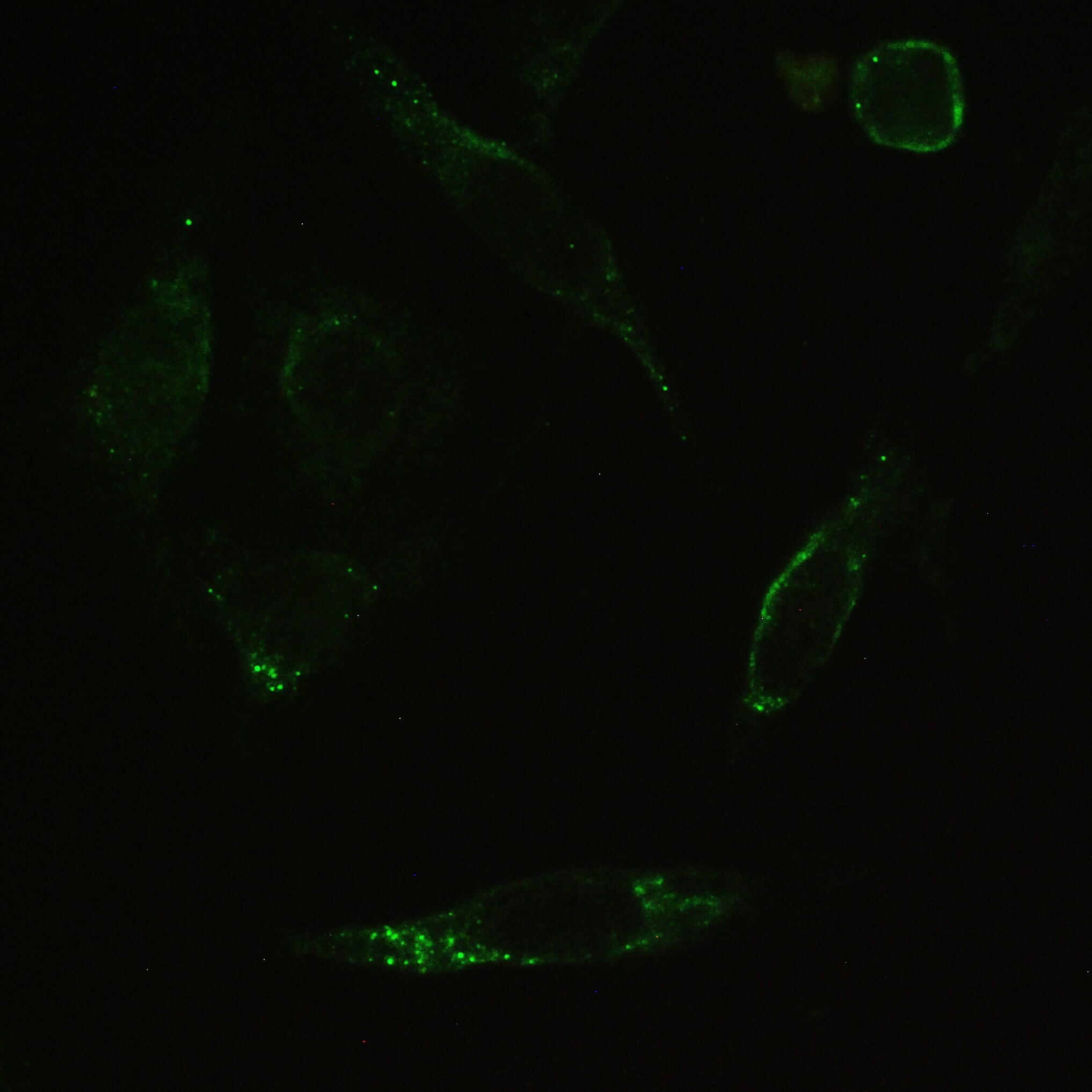
- Immunofluorescent analysis of MYPT1 on U251 cells. Antigen retrieval was performed using citrate buffer (pH6, epitope retrieval solution) for 20 mins. Sample was blocked using 10% goat serum, incubated with MYPT1 polyclonal antibody (Product# PA5-79857) with a dilution of 2 µg/mL (overnight at 4°C). Development was performed using Streptavidin-Biotin-Complex (SABC) with DAB chromogen method.

- Submitted by
- Invitrogen Antibodies (provider)
- Main image

- Experimental details
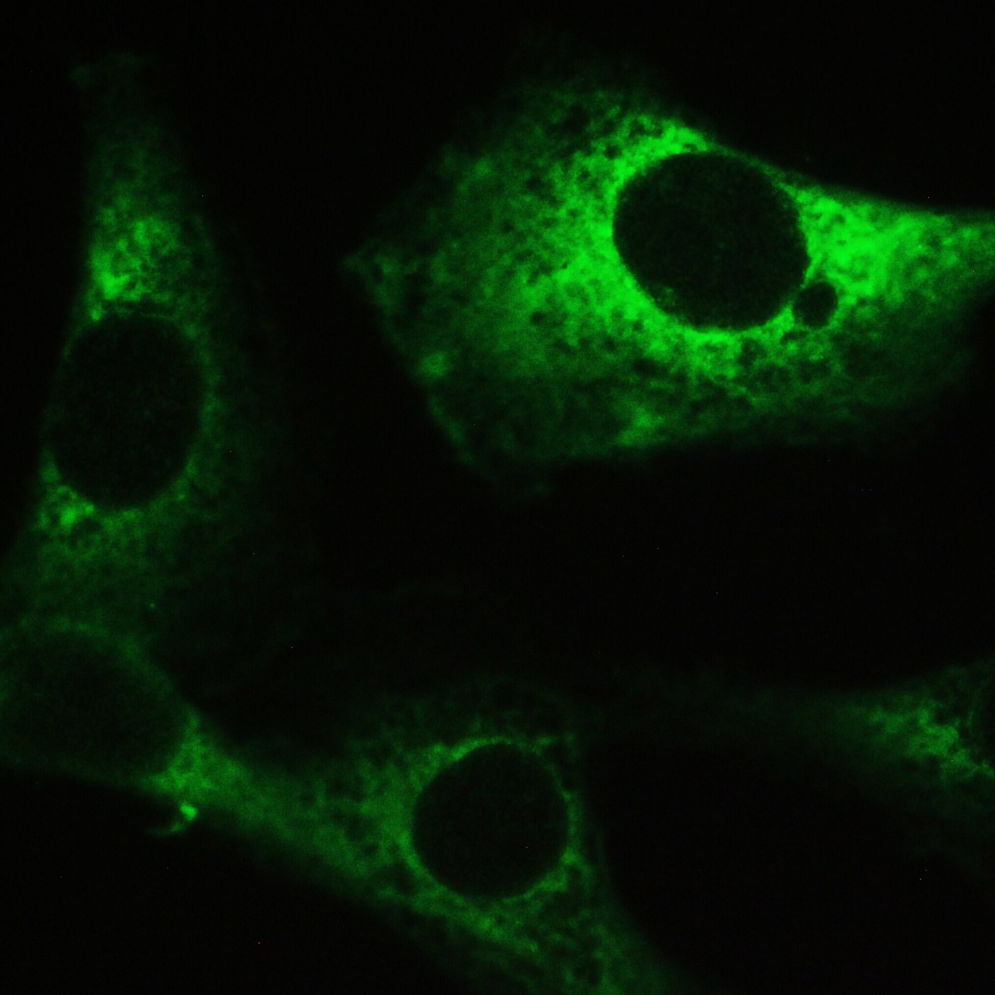
- Immunofluorescent analysis of MYPT1 on U251 cells. Antigen retrieval was performed using citrate buffer (pH6, epitope retrieval solution) for 20 mins. Sample was blocked using 10% goat serum, incubated with MYPT1 polyclonal antibody (Product# PA5-79857) with a dilution of 2 µg/mL (overnight at 4°C). Development was performed using Streptavidin-Biotin-Complex (SABC) with DAB chromogen method.
- Submitted by
- Invitrogen Antibodies (provider)
- Main image
- Experimental details
- Immunofluorescent analysis of MYPT1 on SiHa cells. Antigen retrieval was performed using citrate buffer (pH6, epitope retrieval solution) for 20 mins. Sample was blocked using 10% goat serum, incubated with MYPT1 polyclonal antibody (Product# PA5-79857) with a dilution of 2 µg/mL (overnight at 4°C). Development was performed using Streptavidin-Biotin-Complex (SABC) with DAB chromogen method.
- Submitted by
- Invitrogen Antibodies (provider)
- Main image

- Experimental details
- Immunofluorescent analysis of MYPT1 on SiHa cells. Antigen retrieval was performed using citrate buffer (pH6, epitope retrieval solution) for 20 mins. Sample was blocked using 10% goat serum, incubated with MYPT1 polyclonal antibody (Product# PA5-79857) with a dilution of 2 µg/mL (overnight at 4°C). Development was performed using Streptavidin-Biotin-Complex (SABC) with DAB chromogen method.

- Submitted by
- Invitrogen Antibodies (provider)
- Main image

- Experimental details
- Immunofluorescent analysis of MYPT1 on U251 cells. Antigen retrieval was performed using citrate buffer (pH6, epitope retrieval solution) for 20 mins. Sample was blocked using 10% goat serum, incubated with MYPT1 polyclonal antibody (Product# PA5-79857) with a dilution of 2 µg/mL (overnight at 4°C). Development was performed using Streptavidin-Biotin-Complex (SABC) with DAB chromogen method.

Supportive validation
- Submitted by
- Invitrogen Antibodies (provider)
- Main image
- Experimental details
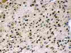
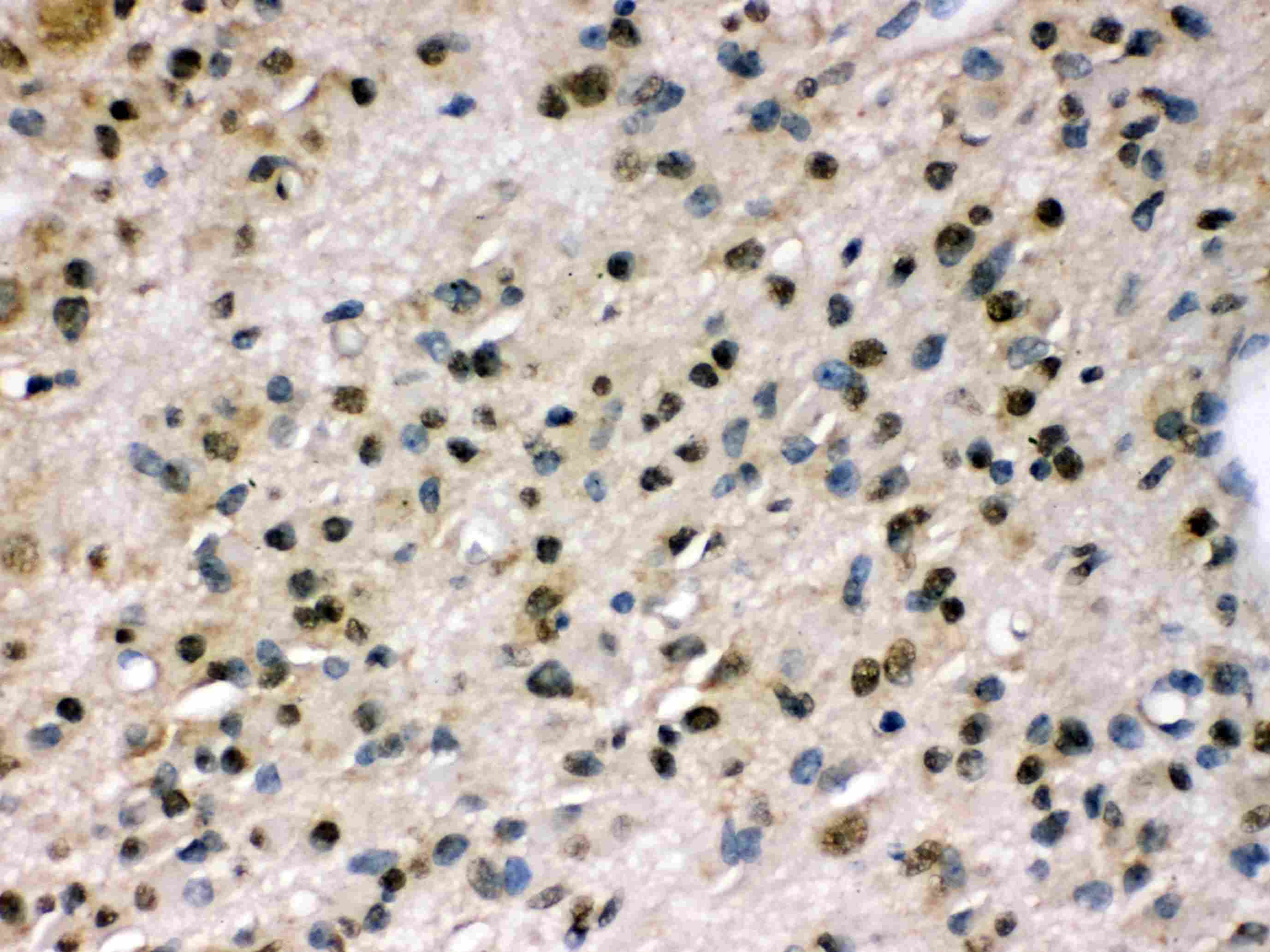
- Immunohistochemistry analysis of MYPT1 on paraffin-embedded human glioma tissue. Antigen retrieval was performed using citrate buffer (pH6, epitope retrieval solution) for 20 mins. Sample was blocked using 10% goat serum, incubated with MYPT1 polyclonal antibody (Product# PA5-79857) with a dilution of 1 µg/mL (overnight at 4°C), and followed by biotinylated goat anti-rabbit IgG (30 minutes at 37°C). Development was performed using Streptavidin-Biotin-Complex (SABC) with DAB chromogen method.
Supportive validation
- Submitted by
- Invitrogen Antibodies (provider)
- Main image

- Experimental details
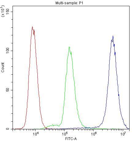
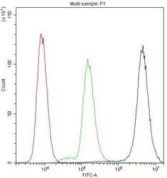
- Flow Cytometry of MYPT1 in HeLa cells (blue line), isotype control rabbit IgG (green line) and unlabeled (red line). Samples were blocked with 10% goat serum, incubated with MYPT1 Polyclonal Antibody (Product # PA5-79857) at a dilution of 1 μg (per 1x10^6 cells), followed by DyLight®488 conjugated goat anti-rabbit IgG (for 30 minutes at 20°C) using 5-10 μg (per 1x10^6 cells) dilution.

- Submitted by
- Invitrogen Antibodies (provider)
- Main image

- Experimental details
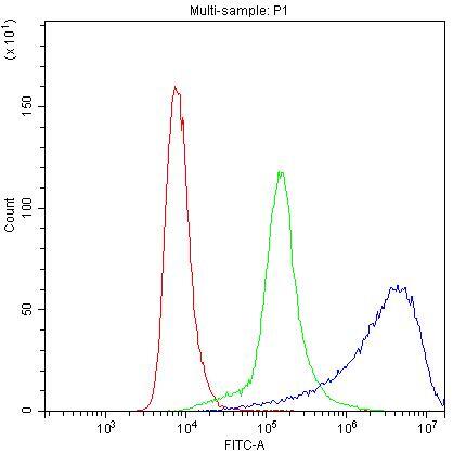
- Flow Cytometry of MYPT1 in U251 cells (blue line), isotype control rabbit IgG (green line) and unlabeled (red line). Samples were blocked with 10% goat serum, incubated with MYPT1 Polyclonal Antibody (Product # PA5-79857) at a dilution of 1 μg (per 1x10^6 cells), followed by DyLight®488 conjugated goat anti-rabbit IgG (for 30 minutes at 20°C) using 5-10 μg (per 1x10^6 cells) dilution.

- Submitted by
- Invitrogen Antibodies (provider)
- Main image
- Experimental details
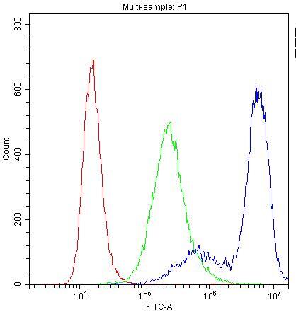
- Flow Cytometry of MYPT1 in SiHa cells (blue line), isotype control rabbit IgG (green line) and unlabeled (red line). Samples were blocked with 10% goat serum, incubated with MYPT1 Polyclonal Antibody (Product # PA5-79857) at a dilution of 1 μg (per 1x10^6 cells), followed by DyLight®488 conjugated goat anti-rabbit IgG (for 30 minutes at 20°C) using 5-10 μg (per 1x10^6 cells) dilution.

- Submitted by
- Invitrogen Antibodies (provider)
- Main image

- Experimental details
- Flow Cytometry of MYPT1 in HeLa cells (blue line), isotype control rabbit IgG (green line) and unlabeled (red line). Samples were blocked with 10% goat serum, incubated with MYPT1 Polyclonal Antibody (Product # PA5-79857) at a dilution of 1 μg (per 1x10^6 cells), followed by DyLight®488 conjugated goat anti-rabbit IgG (for 30 minutes at 20°C) using 5-10 μg (per 1x10^6 cells) dilution.
- Submitted by
- Invitrogen Antibodies (provider)
- Main image

- Experimental details
- Flow Cytometry of MYPT1 in U251 cells (blue line), isotype control rabbit IgG (green line) and unlabeled (red line). Samples were blocked with 10% goat serum, incubated with MYPT1 Polyclonal Antibody (Product # PA5-79857) at a dilution of 1 μg (per 1x10^6 cells), followed by DyLight®488 conjugated goat anti-rabbit IgG (for 30 minutes at 20°C) using 5-10 μg (per 1x10^6 cells) dilution.
- Submitted by
- Invitrogen Antibodies (provider)
- Main image

- Experimental details
- Flow Cytometry of MYPT1 in SiHa cells (blue line), isotype control rabbit IgG (green line) and unlabeled (red line). Samples were blocked with 10% goat serum, incubated with MYPT1 Polyclonal Antibody (Product # PA5-79857) at a dilution of 1 μg (per 1x10^6 cells), followed by DyLight®488 conjugated goat anti-rabbit IgG (for 30 minutes at 20°C) using 5-10 μg (per 1x10^6 cells) dilution.