 Explore
 Validate
 Learn
Learn Western blot
 Immunoprecipitation
Antibody data
- Antibody Data
- Antigen structure
- References [1]
- Comments [0]
- Validations
- Western blot [2]
Submit
Validation data
Reference
Comment
Report error
- Product number
- NB110-38889 - Provider product page

- Provider
- Novus Biologicals
- Proper citation
- Novus Cat#NB110-38889, RRID:AB_791299
- Product name
- Rabbit Polyclonal Myosin Phosphatase Antibody
- Antibody type
- Polyclonal
- Description
- Immunogen affinity purified.
- Reactivity
- Human
- Host
- Rabbit
- Isotype
- IgG
- Vial size
- 100 ul
- Concentration
- 1.0 mg/ml
- Storage
- Store at 4C. Do not freeze.
Submitted references Hypoxia-inducible factors mediate coordinated RhoA-ROCK1 expression and signaling in breast cancer cells.
Gilkes DM, Xiang L, Lee SJ, Chaturvedi P, Hubbi ME, Wirtz D, Semenza GL
Proceedings of the National Academy of Sciences of the United States of America 2014 Jan 21;111(3):E384-93
Proceedings of the National Academy of Sciences of the United States of America 2014 Jan 21;111(3):E384-93
No comments: Submit comment
Supportive validation
- Submitted by
- Novus Biologicals (provider)
- Main image

- Experimental details
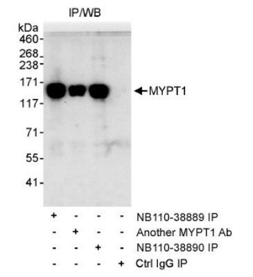
- Western Blot: Myosin Phosphatase Antibody [NB110-38889] - Whole cell lysate (1 mg/IP, 20% IP loaded) from HeLa cells. NB110-38889 used for IP (3 ug/mg lysate). MYPT1 was also immunoprecipitated by other MYPT1 antibody, recognize downstream epitopes.
- Submitted by
- Novus Biologicals (provider)
- Main image
- Experimental details
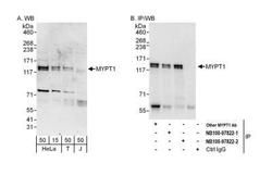
- Western Blot: Myosin Phosphatase Antibody [NB110-38889] - Detection of human MYPT1 by western blot and immunoprecipitation. Samples: Whole cell lysate from HeLa (15 and 50 ug for WB; 1 mg for IP, 20% of IP loaded), HEK293T (T; 50 ug) and Jurkat (J; 50 ug) cells. Antibodies: Affinity purified rabbit anti-MYPT1 antibody NB110-38889 (lot NB110-38889-2) used for WB at 0.4 ug/ml (A) and 1 ug/ml (B) and used for IP at 6 ug/mg lysate (B). MYPT1 was also immunoprecipitated by a previous lot (lot NB110-38889-1) of this antibody and by another rabbit anti-MYPT1 antibody, which recognizes a downstream epitope. Detection: Chemiluminescence with exposure times of 3 minutes (A) and 30 seconds (B).
