 Explore
 Validate
 Learn
Learn Immunocytochemistry
 Immunohistochemistry
Antibody data
- Antibody Data
- Antigen structure
- References [15]
- Comments [0]
- Validations
- Immunocytochemistry [1]
Submit
Validation data
Reference
Comment
Report error
- Product number
- HPA042451 - Provider product page

- Provider
- Atlas Antibodies
- Proper citation
- Atlas Antibodies Cat#HPA042451, RRID:AB_10806239
- Product name
- Anti-SETD2
- Antibody type
- Polyclonal
- Description
- Polyclonal Antibody against Human SETD2, Gene description: SET domain containing 2, Alternative Gene Names: FLJ23184, HIF-1, HYPB, KIAA1732, KMT3A, Validated applications: ICC, IHC, Uniprot ID: Q9BYW2, Storage: Store at +4°C for short term storage. Long time storage is recommended at -20°C.
- Reactivity
- Human
- Host
- Rabbit
- Conjugate
- Unconjugated
- Isotype
- IgG
- Vial size
- 100 µl
- Concentration
- 0.2 mg/ml
- Storage
- Store at +4°C for short term storage. Long time storage is recommended at -20°C.
- Handling
- The antibody solution should be gently mixed before use.
Submitted references Exploring The Prognostic Significance of SET-Domain Containing 2 (SETD2) Expression in Advanced and Castrate-Resistant Prostate Cancer
Multi-omic and single-cell profiling of chromothriptic medulloblastoma reveals genomic and transcriptomic consequences of genome instability.
SETD2 regulates chromatin accessibility and transcription to suppress lung tumorigenesis
SETD2 Loss and ATR Inhibition Synergize to Promote cGAS Signaling and Immunotherapy Response in Renal Cell Carcinoma
SETD2 safeguards the genome against isochromosome formation
SETD2 loss perturbs the kidney cancer epigenetic landscape to promote metastasis and engenders actionable dependencies on histone chaperone complexes
α-TubK40me3 is required for neuronal polarization and migration by promoting microtubule formation
SET Domain Containing 2 Deficiency in Myelodysplastic Syndrome
SETD2 loss sensitizes cells to PI3Kβ and AKT inhibition
SETD2 Haploinsufficiency for Microtubule Methylation Is an Early Driver of Genomic Instability in Renal Cell Carcinoma
SETD2 and histone H3 lysine 36 methylation deficiency in advanced systemic mastocytosis
Methylated α-tubulin antibodies recognize a new microtubule modification on mitotic microtubules
Type II enteropathy-associated T-cell lymphoma features a unique genomic profile with highly recurrent SETD2 alterations
Structure/Function Analysis of Recurrent Mutations in SETD2 Protein Reveals a Critical and Conserved Role for a SET Domain Residue in Maintaining Protein Stability and Histone H3 Lys-36 Trimethylation
SETD2 loss-of-function promotes renal cancer branched evolution through replication stress and impaired DNA repair
Gamallat Y, Felipe Lima J, Seyedi S, Li Q, Rokne J, Alhajj R, Ghosh S, Bismar T
Cancers 2024;16(7):1436
Cancers 2024;16(7):1436
Multi-omic and single-cell profiling of chromothriptic medulloblastoma reveals genomic and transcriptomic consequences of genome instability.
Smirnov P, Przybilla MJ, Simovic-Lorenz M, Parra RG, Susak H, Ratnaparkhe M, Wong JK, Körber V, Mallm JP, Philippos G, Sill M, Kolb T, Kumar R, Casiraghi N, Okonechnikov K, Ghasemi DR, Maaß KK, Pajtler KW, Jauch A, Korshunov A, Höfer T, Zapatka M, Pfister SM, Huber W, Stegle O, Ernst A
Nature communications 2024 Nov 23;15(1):10183
Nature communications 2024 Nov 23;15(1):10183
SETD2 regulates chromatin accessibility and transcription to suppress lung tumorigenesis
Xie Y, Sahin M, Wakamatsu T, Inoue-Yamauchi A, Zhao W, Han S, Nargund A, Yang S, Lyu Y, Hsieh J, Leslie C, Cheng E
JCI Insight 2023;8(4)
JCI Insight 2023;8(4)
SETD2 Loss and ATR Inhibition Synergize to Promote cGAS Signaling and Immunotherapy Response in Renal Cell Carcinoma
Liu X, Zhang Y, McGrail D, Zhang X, Lam T, Hoang A, Hasanov E, Manyam G, Peterson C, Zhu H, Kumar S, Akbani R, Pilie P, Tannir N, Peng G, Jonasch E
Clinical Cancer Research 2023;29(19):4002-4015
Clinical Cancer Research 2023;29(19):4002-4015
SETD2 safeguards the genome against isochromosome formation
Mason F, Kounlavong E, Tebeje A, Dahiya R, Guess T, Khan A, Vlach L, Norris S, Lovejoy C, Dere R, Strahl B, Ohi R, Ly P, Walker C, Rathmell W
Proceedings of the National Academy of Sciences 2023;120(39)
Proceedings of the National Academy of Sciences 2023;120(39)
SETD2 loss perturbs the kidney cancer epigenetic landscape to promote metastasis and engenders actionable dependencies on histone chaperone complexes
Xie Y, Sahin M, Sinha S, Wang Y, Nargund A, Lyu Y, Han S, Dong Y, Hsieh J, Leslie C, Cheng E
Nature Cancer 2022;3(2):188-202
Nature Cancer 2022;3(2):188-202
α-TubK40me3 is required for neuronal polarization and migration by promoting microtubule formation
Xie X, Wang S, Li M, Diao L, Pan X, Chen J, Zou W, Zhang X, Feng W, Bao L
Nature Communications 2021;12(1)
Nature Communications 2021;12(1)
SET Domain Containing 2 Deficiency in Myelodysplastic Syndrome
Li J, Peng Z, Luo F, Chen Y
Frontiers in Genetics 2020;11
Frontiers in Genetics 2020;11
SETD2 loss sensitizes cells to PI3Kβ and AKT inhibition
Terzo E, Lim A, Chytil A, Chiang Y, Farmer L, Gessner K, Walker C, Jansen V, Rathmell W
Oncotarget 2019;10(6):647-659
Oncotarget 2019;10(6):647-659
SETD2 Haploinsufficiency for Microtubule Methylation Is an Early Driver of Genomic Instability in Renal Cell Carcinoma
Chiang Y, Park I, Terzo E, Tripathi D, Mason F, Fahey C, Karki M, Shuster C, Sohn B, Chowdhury P, Powell R, Ohi R, Tsai Y, de Cubas A, Khan A, Davis I, Strahl B, Parker J, Dere R, Walker C, Rathmell W
Cancer Research 2018;78(12):3135-3146
Cancer Research 2018;78(12):3135-3146
SETD2 and histone H3 lysine 36 methylation deficiency in advanced systemic mastocytosis
Martinelli G, Mancini M, De Benedittis C, Rondoni M, Papayannidis C, Manfrini M, Meggendorfer M, Calogero R, Guadagnuolo V, Fontana M, Bavaro L, Padella A, Zago E, Pagano L, Zanotti R, Scaffidi L, Specchia G, Albano F, Merante S, Elena C, Savini P, Gangemi D, Tosi P, Ciceri F, Poletti G, Riccioni L, Morigi F, Delledonne M, Haferlach T, Cavo M, Valent P, Soverini S
Leukemia 2017;32(1):139-148
Leukemia 2017;32(1):139-148
Methylated α-tubulin antibodies recognize a new microtubule modification on mitotic microtubules
Park I, Chowdhury P, Tripathi D, Powell R, Dere R, Terzo E, Rathmell W, Walker C
mAbs 2016;8(8):1590-1597
mAbs 2016;8(8):1590-1597
Type II enteropathy-associated T-cell lymphoma features a unique genomic profile with highly recurrent SETD2 alterations
Roberti A, Dobay M, Bisig B, Vallois D, Boéchat C, Lanitis E, Bouchindhomme B, Parrens M, Bossard C, Quintanilla-Martinez L, Missiaglia E, Gaulard P, de Leval L
Nature Communications 2016;7(1)
Nature Communications 2016;7(1)
Structure/Function Analysis of Recurrent Mutations in SETD2 Protein Reveals a Critical and Conserved Role for a SET Domain Residue in Maintaining Protein Stability and Histone H3 Lys-36 Trimethylation
Hacker K, Fahey C, Shinsky S, Chiang Y, DiFiore J, Jha D, Vo A, Shavit J, Davis I, Strahl B, Rathmell W
Journal of Biological Chemistry 2016;291(40):21283-21295
Journal of Biological Chemistry 2016;291(40):21283-21295
SETD2 loss-of-function promotes renal cancer branched evolution through replication stress and impaired DNA repair
Kanu N, Grönroos E, Martinez P, Burrell R, Yi Goh X, Bartkova J, Maya-Mendoza A, Mistrík M, Rowan A, Patel H, Rabinowitz A, East P, Wilson G, Santos C, McGranahan N, Gulati S, Gerlinger M, Birkbak N, Joshi T, Alexandrov L, Stratton M, Powles T, Matthews N, Bates P, Stewart A, Szallasi Z, Larkin J, Bartek J, Swanton C
Oncogene 2015;34(46):5699-5708
Oncogene 2015;34(46):5699-5708
No comments: Submit comment
Supportive validation
- Submitted by
- Atlas Antibodies (provider)
- Main image
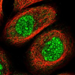
- Experimental details
- Immunofluorescent staining of human cell line A-431 shows localization to nuclear speckles & cytosol.
- Sample type
- Human
