Explore
 Validate
 Learn
Learn Western blot
 ELISA
ELISAAntibody data
- Antibody Data
- Antigen structure
- References [0]
- Comments [0]
- Validations
- Western blot [1]
- Immunohistochemistry [2]
Submit
Validation data
Reference
Comment
Report error
- Product number
- LS-B10609 - Provider product page

- Provider
- LSBio
- Product name
- IHC-plus™ SIPA1 Antibody LS-B10609
- Antibody type
- Polyclonal
- Description
- Affinity purified
- Reactivity
- Human, Mouse, Rat
- Host
- Rabbit
- Isotype
- IgG
- Storage
- Short term: -20°C; Long term: -20°C.
No comments: Submit comment
Enhanced validation
- Submitted by
- LSBio (provider)
- Enhanced method
- Genetic validation
- Main image
- Experimental details
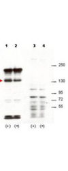
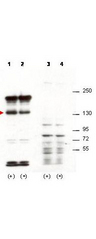
- Anti-Sipa1 Antibody - Western Blot. Western blot of affinity purified anti-Sipa1 antibody shows detection of over-expressed Sipa1 in lysates from mouse 3T3 cells transfected with Sipa1 (lane 1). Endogenous Sipa1 is detected in lane 2, which contains lysate from 3T3 cells mock-transfected with LacZGLB, although at a significantly reduced level compared to transfected cells. Lane 3 and 4 are similar to lanes 1 and 2 except the antibody was preincubated with the immunizing peptide prior to reaction with the membrane. The identity of the higher and lower molecular weight bands is unknown. The band at ~130 kD, indicated by the arrowhead, corresponds to recombinant Sipa1. Primary antibody was used at 1:1250. Personal communication, H. Yang, L. Lukes and K. Hunter, NCI, Bethesda, MD.
Enhanced validation
- Submitted by
- LSBio (provider)
- Enhanced method
- Genetic validation
- Main image

- Experimental details
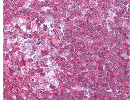
- Anti-Sipa1 Antibody - Immunohistochemistry. affinity purified anti-Sipa1 antibody was used at 1.25 ug/ml to detect signal in a variety of tissues including multi-human, multi-brain and multi-cancer slides. This image shows moderate to strong positive staining of lymphocytes within human tonsil at 40X. Tissue was formalin-fixed and paraffin embedded. The image shows localization of the antibody as the precipitated red signal, with a hematoxylin purple nuclear counterstain.

- Submitted by
- LSBio (provider)
- Enhanced method
- Genetic validation
- Main image
- Experimental details
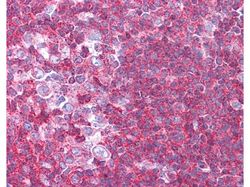
- Anti-Sipa1 Antibody - Immunohistochemistry. affinity purified anti-Sipa1 antibody was used at 1.25 ug/ml to detect signal in a variety of tissues including multi-human, multi-brain and multi-cancer slides. This image shows moderate to strong positive staining of lymphocytes within human tonsil at 40X. Tissue was formalin-fixed and paraffin embedded. The image shows localization of the antibody as the precipitated red signal, with a hematoxylin purple nuclear counterstain.