 Explore
 Validate
 Learn
Learn41-0200
antibody from Invitrogen Antibodies
Targeting: NT5E
CALJA, CD73, eN, eNT, NT5
 Western blot
 Immunocytochemistry
 Immunoprecipitation Immunohistochemistry Flow cytometry Other assay
Antibody data
- Antibody Data
- Antigen structure
- References [10]
- Comments [0]
- Validations
- Immunocytochemistry [2]
- Flow cytometry [2]
- Other assay [2]
Submit
Validation data
Reference
Comment
Report error
- Product number
- 41-0200 - Provider product page

- Provider
- Invitrogen Antibodies
- Product name
- CD73 Monoclonal Antibody (7G2)
- Antibody type
- Monoclonal
- Antigen
- Purifed from natural sources
- Reactivity
- Human
- Host
- Mouse
- Isotype
- IgG
- Antibody clone number
- 7G2
- Vial size
- 100 μg
- Concentration
- 0.5 mg/mL
- Storage
- -20°C
Submitted references Mitochondrial Transfer of Wharton's Jelly Mesenchymal Stem Cells Eliminates Mutation Burden and Rescues Mitochondrial Bioenergetics in Rotenone-Stressed MELAS Fibroblasts.
Extracellular Vesicles from Adipose-Derived Mesenchymal Stem/Stromal Cells Accelerate Migration and Activate AKT Pathway in Human Keratinocytes and Fibroblasts Independently of miR-205 Activity.
Expression regulation and functional analysis of RGS2 and RGS4 in adipogenic and osteogenic differentiation of human mesenchymal stem cells.
Characterization of Tunneling Nanotubes in Wharton's jelly Mesenchymal Stem Cells. An Intercellular Exchange of Components between Neighboring Cells.
Blood-based markers of efficacy and resistance to cetuximab treatment in metastatic colorectal cancer: results from CALGB 80203 (Alliance).
Dengue virus type 2 modulates endothelial barrier function through CD73.
Mesenchymal markers on human adipose stem/progenitor cells.
Morphological and immunocytochemical characteristics indicate the yield of early progenitors and represent a quality control for human mesenchymal stem cell culturing.
Production and characterization of monoclonal antibodies to the glycosyl phosphatidylinositol-anchored lymphocyte differentiation antigen ecto-5'-nucleotidase (CD73).
Production and characterization of monoclonal antibodies to the glycosyl phosphatidylinositol-anchored lymphocyte differentiation antigen ecto-5'-nucleotidase (CD73).
Lin TK, Chen SD, Chuang YC, Lan MY, Chuang JH, Wang PW, Hsu TY, Wang FS, Tsai MH, Huang ST, Wang XW, Tsai PC, Lin HY, Liou CW
Oxidative medicine and cellular longevity 2019;2019:9537504
Oxidative medicine and cellular longevity 2019;2019:9537504
Extracellular Vesicles from Adipose-Derived Mesenchymal Stem/Stromal Cells Accelerate Migration and Activate AKT Pathway in Human Keratinocytes and Fibroblasts Independently of miR-205 Activity.
Ferreira ADF, Cunha PDS, Carregal VM, da Silva PC, de Miranda MC, Kunrath-Lima M, de Melo MIA, Faraco CCF, Barbosa JL, Frezard F, Resende V, Rodrigues MA, de Goes AM, Gomes DA
Stem cells international 2017;2017:9841035
Stem cells international 2017;2017:9841035
Expression regulation and functional analysis of RGS2 and RGS4 in adipogenic and osteogenic differentiation of human mesenchymal stem cells.
Madrigal A, Tan L, Zhao Y
Biological research 2017 Dec 26;50(1):43
Biological research 2017 Dec 26;50(1):43
Characterization of Tunneling Nanotubes in Wharton's jelly Mesenchymal Stem Cells. An Intercellular Exchange of Components between Neighboring Cells.
Sanchez V, Villalba N, Fiore L, Luzzani C, Miriuka S, Boveris A, Gelpi RJ, Brusco A, Poderoso JJ
Stem cell reviews and reports 2017 Aug;13(4):491-498
Stem cell reviews and reports 2017 Aug;13(4):491-498
Blood-based markers of efficacy and resistance to cetuximab treatment in metastatic colorectal cancer: results from CALGB 80203 (Alliance).
Hatch AJ, Sibley AB, Starr MD, Brady JC, Jiang C, Jia J, Bowers DL, Pang H, Owzar K, Niedzwiecki D, Innocenti F, Venook AP, Hurwitz HI, Nixon AB, Alliance for Clinical Trials in Oncology
Cancer medicine 2016 Sep;5(9):2249-60
Cancer medicine 2016 Sep;5(9):2249-60
Dengue virus type 2 modulates endothelial barrier function through CD73.
Patkar C, Giaya K, Libraty DH
The American journal of tropical medicine and hygiene 2013 Jan;88(1):89-94
The American journal of tropical medicine and hygiene 2013 Jan;88(1):89-94
Mesenchymal markers on human adipose stem/progenitor cells.
Zimmerlin L, Donnenberg VS, Rubin JP, Donnenberg AD
Cytometry. Part A : the journal of the International Society for Analytical Cytology 2013 Jan;83(1):134-40
Cytometry. Part A : the journal of the International Society for Analytical Cytology 2013 Jan;83(1):134-40
Morphological and immunocytochemical characteristics indicate the yield of early progenitors and represent a quality control for human mesenchymal stem cell culturing.
Haasters F, Prall WC, Anz D, Bourquin C, Pautke C, Endres S, Mutschler W, Docheva D, Schieker M
Journal of anatomy 2009 May;214(5):759-67
Journal of anatomy 2009 May;214(5):759-67
Production and characterization of monoclonal antibodies to the glycosyl phosphatidylinositol-anchored lymphocyte differentiation antigen ecto-5'-nucleotidase (CD73).
Thomson LF, Ruedi JM, Glass A, Moldenhauer G, Moller P, Low MG, Klemens MR, Massaia M, Lucas AH
Tissue antigens 1990 Jan;35(1):9-19
Tissue antigens 1990 Jan;35(1):9-19
No comments: Submit comment
Supportive validation
- Submitted by
- Invitrogen Antibodies (provider)
- Main image

- Experimental details
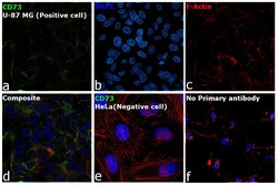
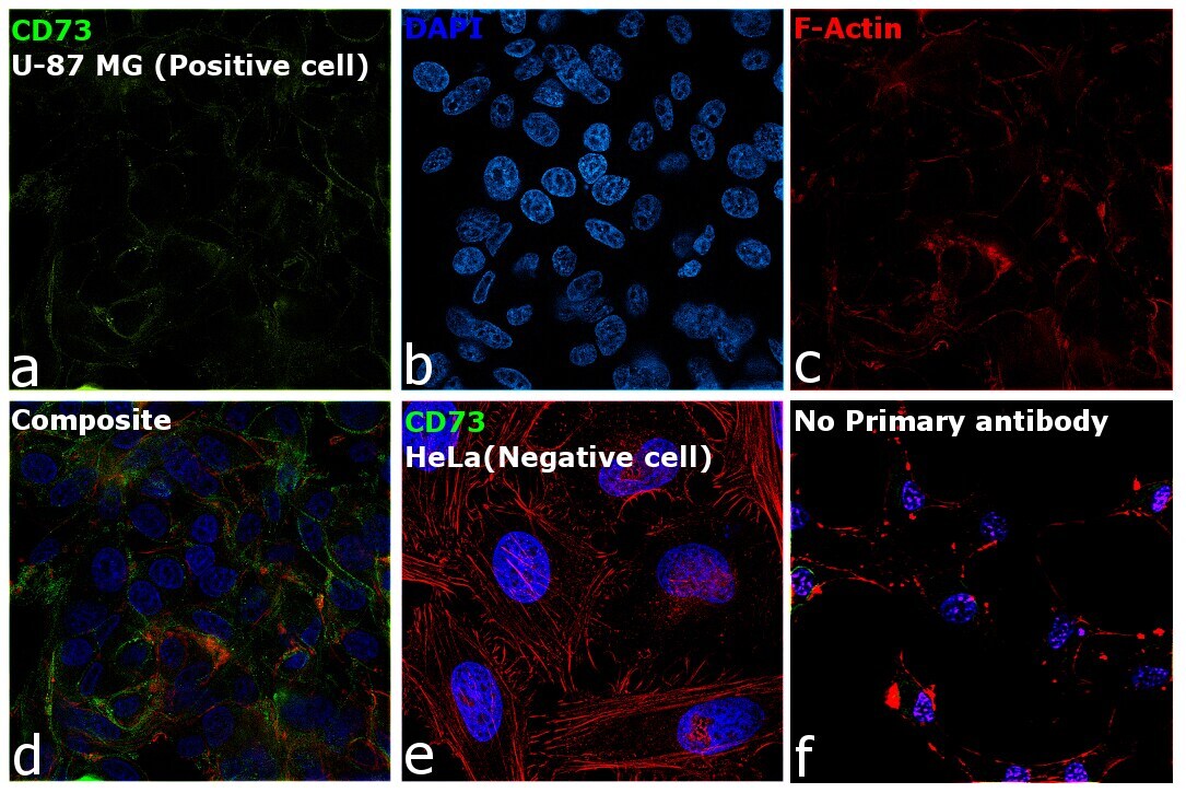
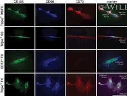
- Immunofluorescence analysis of CD73 was performed using 70% confluent log phase U-87 MG cells. The cells were fixed with 4% paraformaldehyde for 15 minutes, permeabilized with 0.1% Triton™ X-100 for 10 minutes, and blocked with 2% BSA for 45 minutes at room temperature. The cells were labeled with CD73 Monoclonal Antibody (7G2) (Product # 41-0200) at a concentration of 1 µg/mL in 0.1% BSA, incubated at 4 degree celsius overnight and then labeled with Goat anti-Mouse IgG (H+L) Superclonal™ Recombinant Secondary Antibody, Alexa Fluor® 488 conjugate (Product # A28175), (1:2000 dilution), for 45 minutes at room temperature (Panel a: Green). Nuclei (Panel b: Blue) were stained with ProLong™ Diamond Antifade Mountant with DAPI (Product # P36962). F-actin (Panel c: Red) was stained with Rhodamine Phalloidin (Product # R415, 1:300 dilution). Panel d represents the merged image showing membrane localization. Panel e represents HeLa cells showing no expression of CD73. Panel f represents control cells with no primary antibody to assess background. The images were captured at 60X magnification.

- Submitted by
- Invitrogen Antibodies (provider)
- Main image
- Experimental details
- Immunofluorescence analysis of CD73 was performed using 70% confluent log phase U-87 MG cells. The cells were fixed with 4% paraformaldehyde for 15 minutes, permeabilized with 0.1% Triton™ X-100 for 10 minutes, and blocked with 2% BSA for 45 minutes at room temperature. The cells were labeled with CD73 Monoclonal Antibody (7G2) (Product # 41-0200) at a concentration of 1 µg/mL in 0.1% BSA, incubated at 4 degree celsius overnight and then labeled with Goat anti-Mouse IgG (H+L) Superclonal™ Recombinant Secondary Antibody, Alexa Fluor® 488 conjugate (Product # A28175), (1:2000 dilution), for 45 minutes at room temperature (Panel a: Green). Nuclei (Panel b: Blue) were stained with ProLong™ Diamond Antifade Mountant with DAPI (Product # P36962). F-actin (Panel c: Red) was stained with Rhodamine Phalloidin (Product # R415, 1:300 dilution). Panel d represents the merged image showing membrane localization. Panel e represents HeLa cells showing no expression of CD73. Panel f represents control cells with no primary antibody to assess background. The images were captured at 60X magnification.
Supportive validation
- Submitted by
- Invitrogen Antibodies (provider)
- Main image
- Experimental details
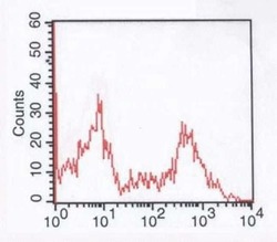
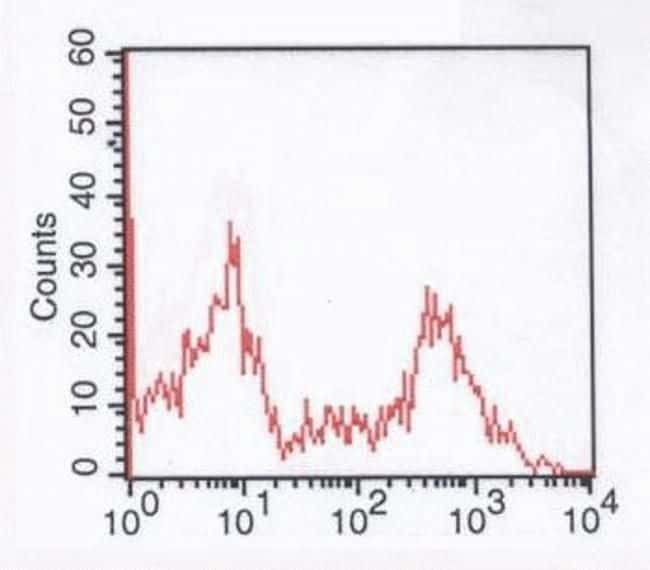
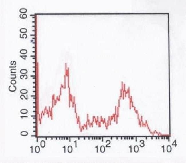
- Human peripheral blood lymphocytes stained with Mouse anti-CD73 monoclonal antibody (Product # 41-0200).
- Submitted by
- Invitrogen Antibodies (provider)
- Main image

- Experimental details
- Human peripheral blood lymphocytes stained with Mouse anti-CD73 monoclonal antibody (Product # 41-0200).
Supportive validation
- Submitted by
- Invitrogen Antibodies (provider)
- Main image
- Experimental details
- NULL

- Submitted by
- Invitrogen Antibodies (provider)
- Main image
- Experimental details
- NULL
