Explore
 Validate
 Learn
Learn Western blot
 Immunohistochemistry
Antibody data
- Antibody Data
- Antigen structure
- References [1]
- Comments [0]
- Validations
- Immunohistochemistry [1]
- Other assay [1]
Submit
Validation data
Reference
Comment
Report error
- Product number
- PA5-18189 - Provider product page

- Provider
- Invitrogen Antibodies
- Product name
- FBXW2 Polyclonal Antibody
- Antibody type
- Polyclonal
- Antigen
- Synthetic peptide
- Description
- This antibody is predicted to react with bovine, canine and porcine based on sequence homology. This antibody is tested in Peptide ELISA: antibody detection limit dilution 128,000.
- Reactivity
- Human, Mouse, Rat
- Host
- Goat
- Isotype
- IgG
- Vial size
- 100 μg
- Concentration
- 0.5 mg/mL
- Storage
- -20°C, Avoid Freeze/Thaw Cycles
Submitted references E3 Ligase FBXW2 Is a New Therapeutic Target in Obesity and Atherosclerosis.
Wang C, Xu W, Chao Y, Liang M, Zhang F, Huang K
Advanced science (Weinheim, Baden-Wurttemberg, Germany) 2020 Oct;7(20):2001800
Advanced science (Weinheim, Baden-Wurttemberg, Germany) 2020 Oct;7(20):2001800
No comments: Submit comment
Supportive validation
- Submitted by
- Invitrogen Antibodies (provider)
- Main image
- Experimental details
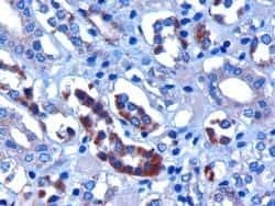
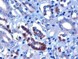
- Immunohistochemistry (PFA fixed) analysis of FBXW2 using FBXW2 Polyclonal Antibody (Product # PA5-18189) (3 µg/mL) in staining of paraffin embedded Human Kidney. Microwaved antigen retrieval with citrate buffer pH 6, HRP-staining.
Supportive validation
- Submitted by
- Invitrogen Antibodies (provider)
- Main image
- Experimental details
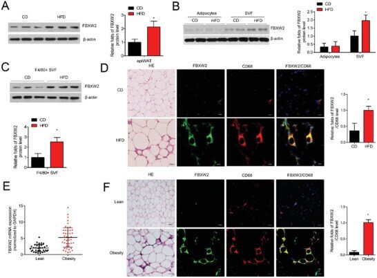
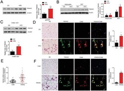
- Figure 1 FBXW2 expression in macrophages is upregulated from obese mice and humans. Eight-week-old C57BL/6J mice were fed either chow diet (CD) or high fat diet (HFD) for 12 weeks. A) FBXW2 expression in epiWAT was determined by western blot assay ( n = 8). B) Cell extracts from isolated adipocytes and SVFs in epiWAT were subject to western blot for the expression of FBXW2 ( n = 5). C) The protein level of FBXW2 in F4/80 + macrophages sorted from SVFs by western blot ( n = 5). D) Representative H&E staining of epiWAT, and the immunofluorescence images for CD68 (red) and FBXW2 (green) expression in epiWAT. Nuclei staining by DAPI (blue). Scale bars, 20 um. E) CD14 + macrophages were isolated from visceral adipose tissue of lean ( n = 33) and obese ( n = 42) subjects. The mRNA level of FBXW2 was tested by real-time qPCR assay. F) Representative H&E staining of visceral adipose tissues from lean and obese individuals, and the expressions of CD68 (red) and FBXW2 (green) were detected by immunofluorescence staining. Nuclei staining by DAPI (blue). Scale bars, 20 um. The data are shown as the mean +- SEM. * p < 0.05 by Student's t test or ANOVA with the post-hoc test.