Explore
 Validate
 Learn
Learn Western blot
 Immunocytochemistry
Antibody data
- Antibody Data
- Antigen structure
- References [1]
- Comments [0]
- Validations
- Immunocytochemistry [2]
- Immunohistochemistry [2]
- Other assay [2]
Submit
Validation data
Reference
Comment
Report error
- Product number
- PA5-14607 - Provider product page

- Provider
- Invitrogen Antibodies
- Product name
- EphB2 Polyclonal Antibody
- Antibody type
- Polyclonal
- Antigen
- Synthetic peptide
- Reactivity
- Human
- Host
- Rabbit
- Isotype
- IgG
- Vial size
- 400 μL
- Concentration
- 2 mg/mL
- Storage
- Store at 4°C short term. For long term storage, store at -20°C, avoiding freeze/thaw cycles.
Submitted references Expression of ID4 protein in breast cancer cells induces reprogramming of tumour-associated macrophages.
Donzelli S, Milano E, Pruszko M, Sacconi A, Masciarelli S, Iosue I, Melucci E, Gallo E, Terrenato I, Mottolese M, Zylicz M, Zylicz A, Fazi F, Blandino G, Fontemaggi G
Breast cancer research : BCR 2018 Jun 19;20(1):59
Breast cancer research : BCR 2018 Jun 19;20(1):59
No comments: Submit comment
Supportive validation
- Submitted by
- Invitrogen Antibodies (provider)
- Main image

- Experimental details
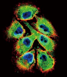
- Immunofluorescent analysis of A375 cells using an EphB2 polyclonal antibody (Product # PA5-14607) at a dilution of 1:10-50, followed by a fluor-conjugated goat anti-rabbit secondary antibody (green). Actin filaments were stained with dye-conjugated phalloidin (red). Nuclei were stained with DAPI (blue).

- Submitted by
- Invitrogen Antibodies (provider)
- Main image
- Experimental details
- Immunocytochemistry analysis of EphB2 in A375 cells. Samples were incubated in EphB2 polyclonal antibody (Product # PA5-14607) followed by Alexa Fluor 488-conjugated goat anti-rabbit lgG (green). Actin filaments have been labeled with Alexa Fluor 555 phalloidin (red). DAPI was used to stain the cell nuclear (blue).

Supportive validation
- Submitted by
- Invitrogen Antibodies (provider)
- Main image
- Experimental details
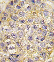
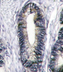
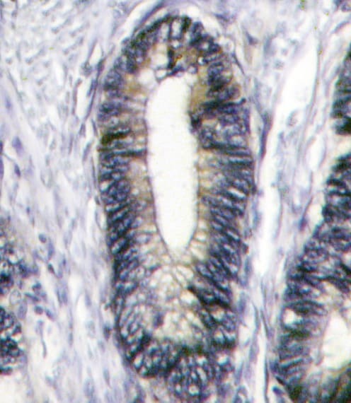
- Immunohistochemistry analysis of EphB2 in formalin-fixed and paraffin-embedded human breast carcinoma tissue. Samples were incubated with EphB2 polyclonal antibody (Product # PA5-14607) which was peroxidase-conjugated to the secondary antibody, followed by DAB staining. This data demonstrates the use of this antibody for immunohistochemistry; clinical relevance has not been evaluated.

- Submitted by
- Invitrogen Antibodies (provider)
- Main image
- Experimental details
- Immunohistochemistry analysis of EphB2 in formalin-fixed and paraffin-embedded human breast carcinoma. Samples were incubated with EphB2 polyclonal antibody (Product # PA5-14607) which was peroxidase-conjugated to the secondary antibody, followed by DAB staining. This data demonstrates the use of this antibody for immunohistochemistry; clinical relevance has not been evaluated.
Supportive validation
- Submitted by
- Invitrogen Antibodies (provider)
- Main image

- Experimental details
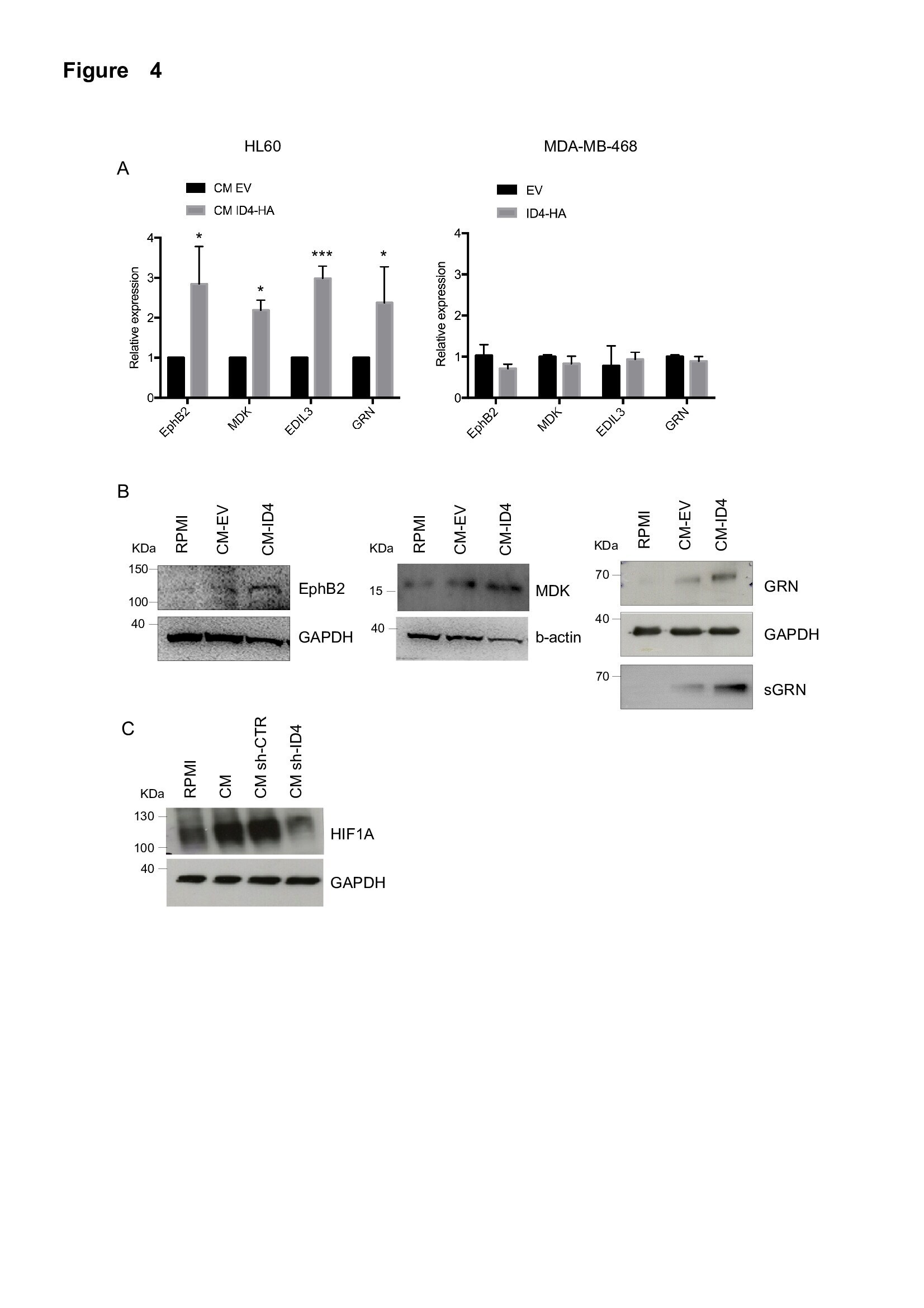
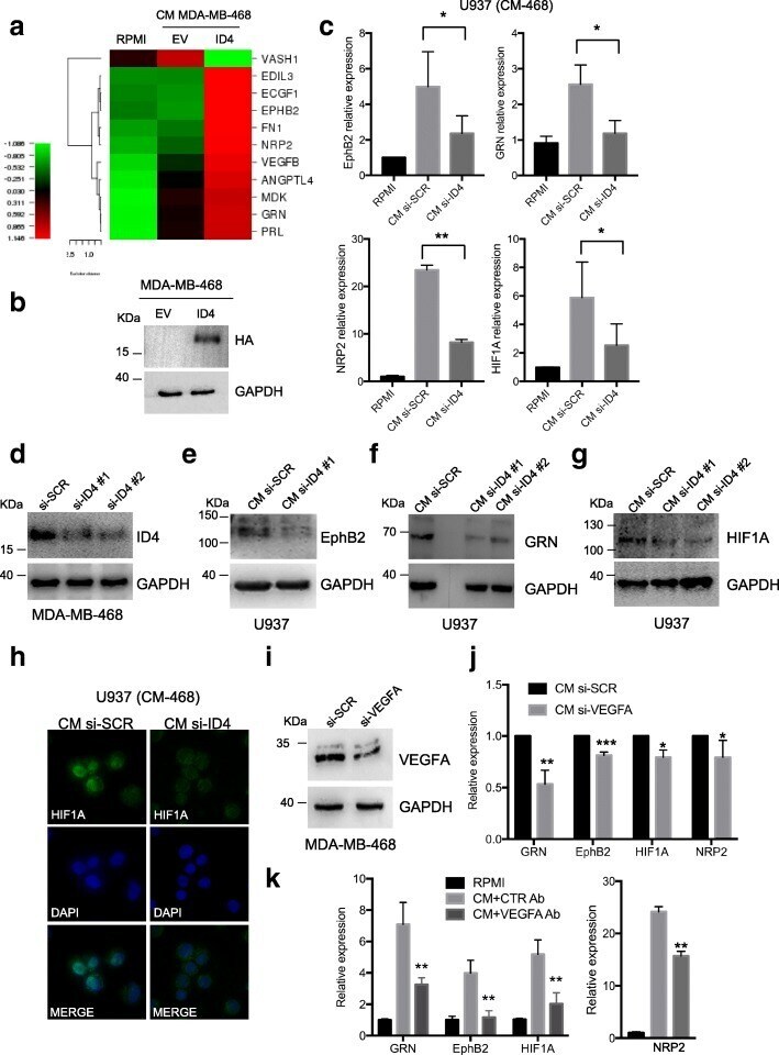
- Fig. 3 Inhibitor of differentiation 4 (ID4) expression in breast cancer cells leads to the activation of an angiogenic programme in macrophages. a Expression matrix representing a panel of angiogenic factors evaluated using TaqMan Low-Density Arrays (TLDA) in macrophages obtained by 1,25-dihydroxyvitamin D 3 (VitD3)-mediated differentiation of HL60 cells and subsequently cultured in RPMI medium or in conditioned media (CM) from control (EV) or ID4-overexpressing (ID4) MDA-MB-468 breast cancer cells. b Western blot showing ID4-HA overexpression in MDA-MB-468 cells. c Selected genes modulated in the arrays were evaluated by RT-qPCR in macrophages obtained from VitD3-mediated differentiation of U937 cells and subsequently cultivated in RPMI medium (CTR) or in CM from control (CM si-SCR) or ID4-depleted (CM si-ID4) MDA-MB-468 cells. d Western blot analysis showing the level of ID4 protein after transfection of the indicated small interfering RNAs (siRNAs) in MDA-MB-468 cells. e - g Western blot analysis of ephrin B2 (EphB2), granulin (GRN) and hypoxia-inducible factor (HIF)-1A proteins in differentiated U937 cells cultured in CM si-SCR or CM si-ID4 from MDA-MB-468 cells. h Immunofluorescence analysis of HIF-1A protein performed in differentiated U937 cells cultured in the presence of CM si-SCR or CM si-ID4 from MDA-MB-468 cells. i Western blotting showing the efficiency of vascular endothelial growth factor A (VEGFA) depletion by siRNA transfection in MDA-MB-468 cells used to pre
- Submitted by
- Invitrogen Antibodies (provider)
- Main image
- Experimental details
- Additional file 7: Figure S4 a Modulation of selected genes modulated in the TLDA was validated by RT-qPCR in differentiated HL60 cells cultured in CM from ID4-overexpressing (CM ID4-HA) or control (CM EV) MDA-MB-468 cells (left panel). The same transcripts were analysed in MDA-MB-468 cells transfected with ID4-HA expression vector (ID4-HA) or control empty vector (EV) (right panel). b Expression of EphB2, MDK and GRN protein evaluated by Western blotting on lysates from differentiated HL60 cells cultured as in ( a ); secreted GRN (sGRN) was evaluated on CM from differentiated HL60 cells in the same conditions. c HIF1A protein expression evaluated by Western blotting in differentiated U937 cells cultured in RPMI medium or in CM from SKBR3 cells stably interfered for ID4 expression (sh-ID4) or control cells (sh-CTR). (PDF 1320 kb)