Explore
 Validate
 Learn
Learn Western blot
 Immunocytochemistry
Antibody data
- Antibody Data
- Antigen structure
- References [1]
- Comments [0]
- Validations
- Immunocytochemistry [1]
- Immunohistochemistry [1]
Submit
Validation data
Reference
Comment
Report error
- Product number
- HPA057052 - Provider product page

- Provider
- Atlas Antibodies
- Proper citation
- Atlas Antibodies Cat#HPA057052, RRID:AB_2683322
- Product name
- Anti-LIPA
- Antibody type
- Polyclonal
- Description
- Polyclonal Antibody against Human LIPA, Gene description: lipase A, lysosomal acid, cholesterol esterase, Alternative Gene Names: CESD, LAL, Validated applications: ICC, IHC, WB, Uniprot ID: P38571, Storage: Store at +4°C for short term storage. Long time storage is recommended at -20°C.
- Reactivity
- Human
- Host
- Rabbit
- Conjugate
- Unconjugated
- Isotype
- IgG
- Vial size
- 100 µl
- Concentration
- 0.1 mg/ml
- Storage
- Store at +4°C for short term storage. Long time storage is recommended at -20°C.
- Handling
- The antibody solution should be gently mixed before use.
Submitted references Therapeutic efficacy of rscAAVrh74.miniCMV.LIPA gene therapy in a mouse model of lysosomal acid lipase deficiency
Lam P, Ashbrook A, Zygmunt D, Yan C, Du H, Martin P
Molecular Therapy - Methods & Clinical Development 2022;26
Molecular Therapy - Methods & Clinical Development 2022;26
No comments: Submit comment
Supportive validation
- Submitted by
- Atlas Antibodies (provider)
- Main image

- Experimental details
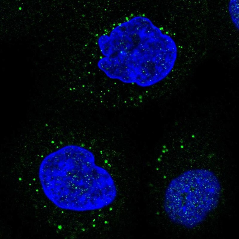
- Immunofluorescent staining of human cell line A-431 shows localization to vesicles.
- Sample type
- Human
Supportive validation
- Submitted by
- Atlas Antibodies (provider)
- Enhanced method
- Orthogonal validation
- Main image
- Experimental details
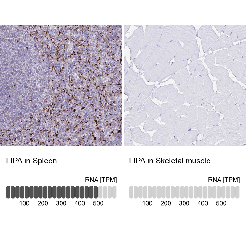
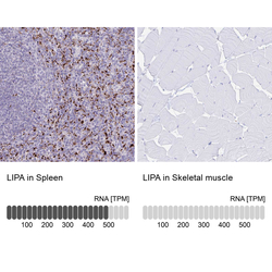
- Immunohistochemistry analysis in human spleen and skeletal muscle tissues using HPA057052 antibody. Corresponding LIPA RNA-seq data are presented for the same tissues.
- Sample type
- Human
- Protocol
- Protocol